. 2010 Aug;6(8):2979-90.
doi: 10.1016/j.actbio.2010.02.035. Epub 2010 Feb 25.

A novel platform for in situ investigation of cells and tissues under mechanical strain

Affiliations

A novel platform for in situ investigation of cells and tissues under mechanical strain

W W Ahmed et al. Acta Biomater. 2010 Aug.

Abstract

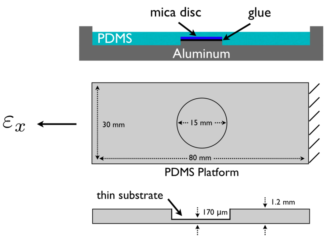
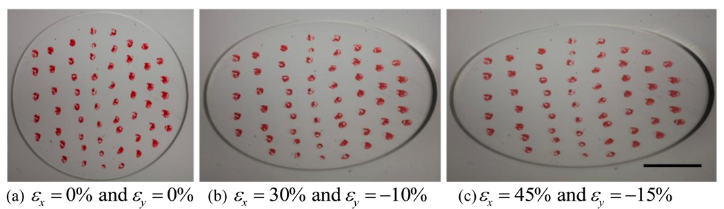
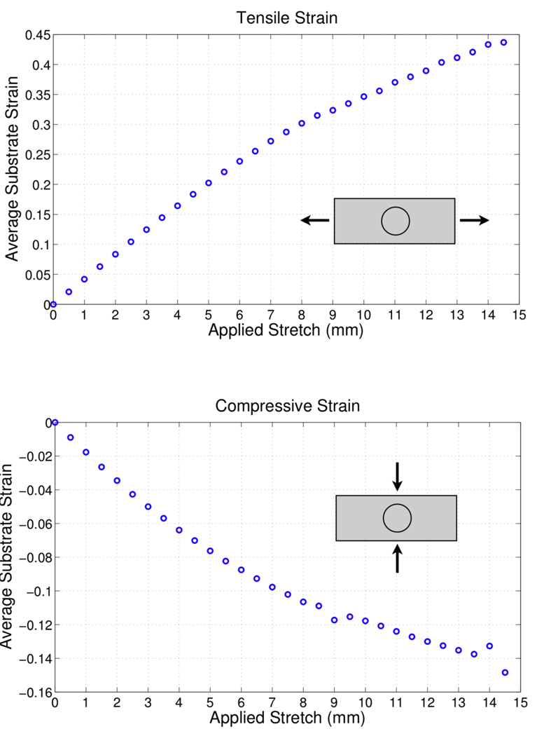
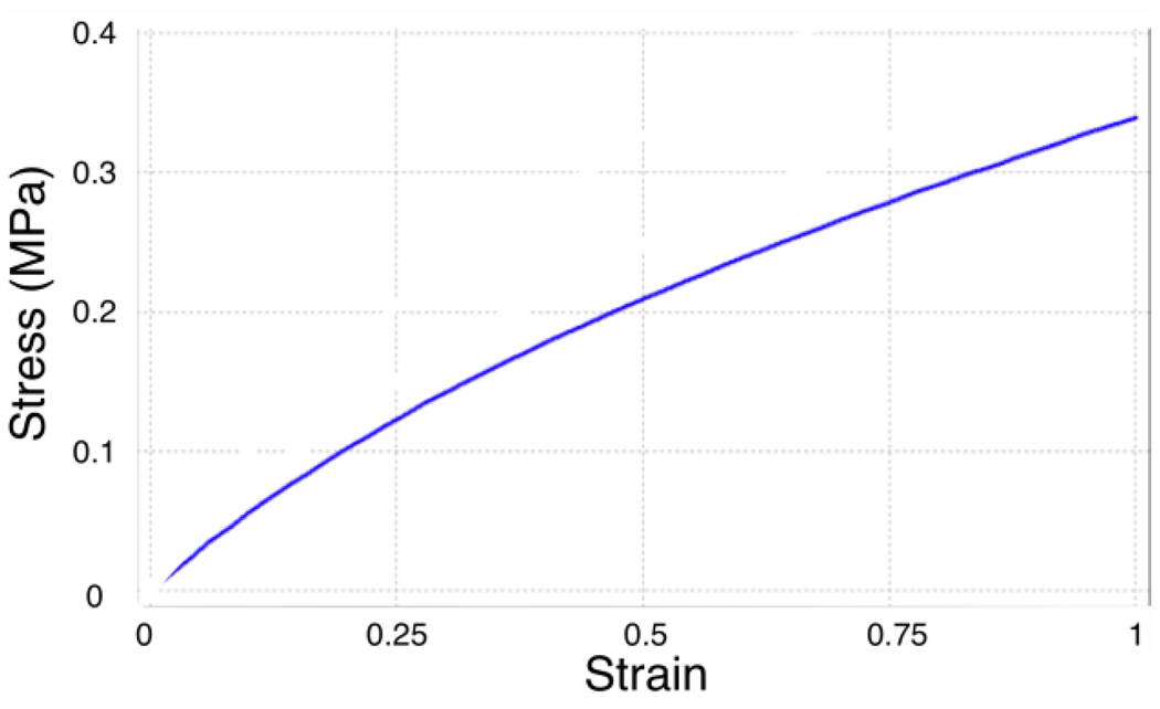
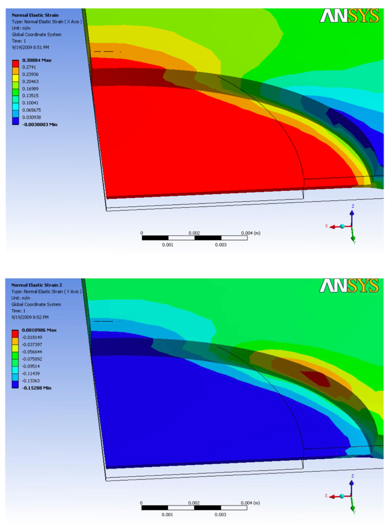
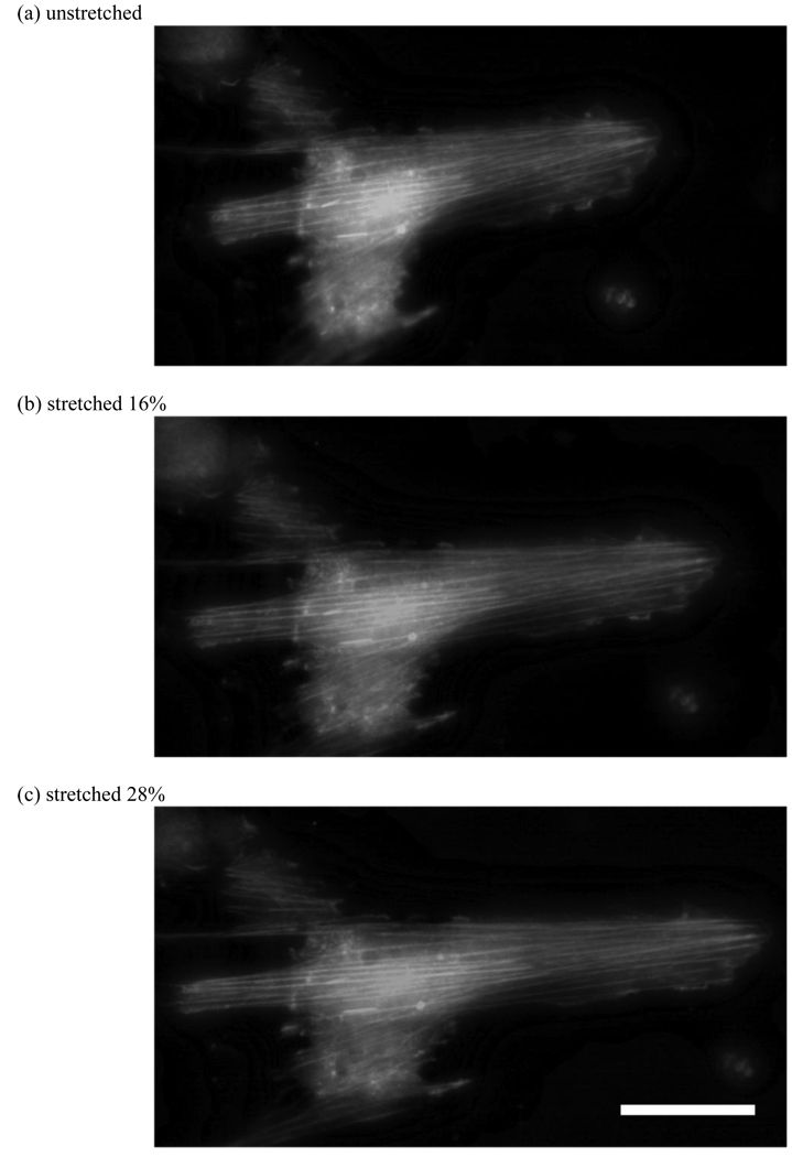
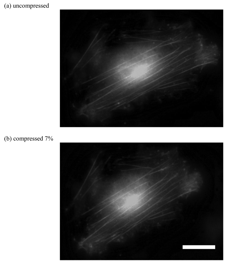
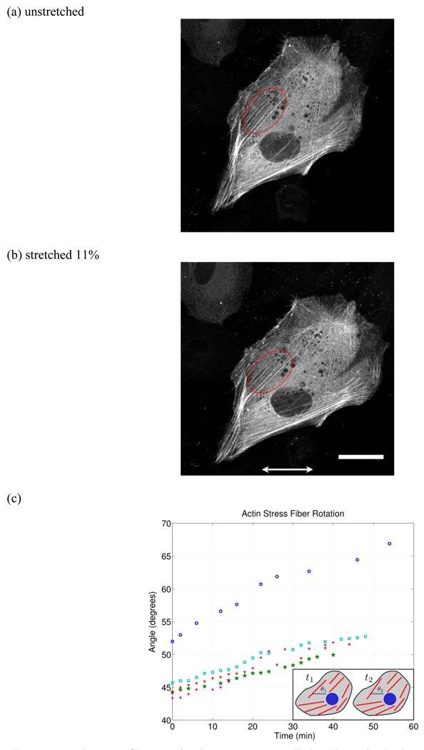
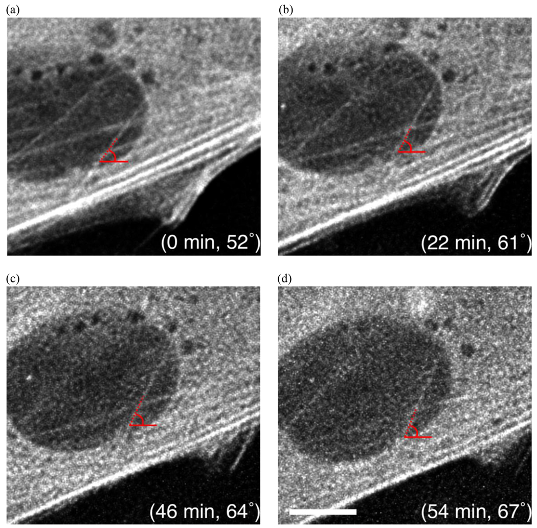
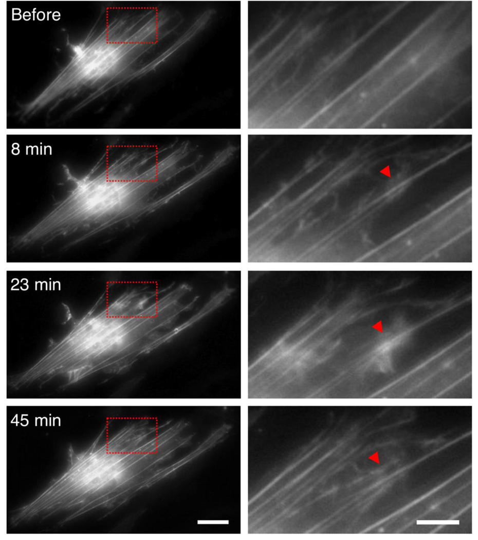
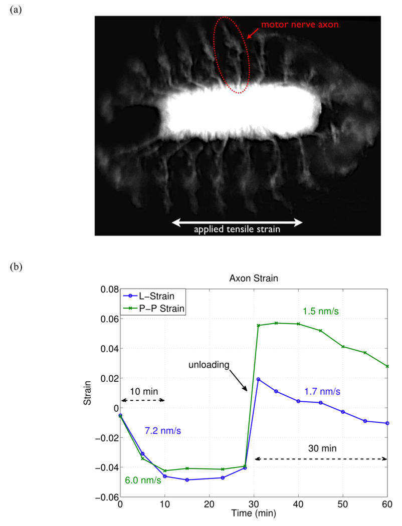
The mechanical micro-environment influences cellular responses such as migration, proliferation, differentiation and apoptosis. Cells are subjected to mechanical stretching in vivo, e.g., epithelial cells during embryogenesis. Current methodologies do not allow high-resolution in situ observation of cells and tissues under applied strain, which may reveal intracellular dynamics and the origin of cell mechanosensitivity. A novel polydimethylsiloxane substrate was developed, capable of applying tensile and compressive strain (up to 45%) to cells and tissues while allowing in situ observation with high-resolution optics. The strain field of the substrate was characterized experimentally using digital image correlation, and the deformation was modeled by the finite element method, using a Mooney-Rivlin hyperelastic constitutive relation. The substrate strain was found to be uniform for >95% of the substrate area. As a demonstration of the system, mechanical strain was applied to single fibroblasts transfected with GFP-actin and whole transgenic Drosophila embryos expressing GFP in all neurons during live imaging. Three observations of biological responses due to applied strain are reported: (1) dynamic rotation of intact actin stress fibers in fibroblasts; (2) lamellipodia activity and actin polymerization in fibroblasts; (3) active axonal contraction in Drosophila embryo motor neurons. The novel platform may serve as an important tool in studying the mechanoresponse of cells and tissues, including whole embryos.


Figures

Figure 1
Figure 1
Schematic diagrams of the aluminum mold and the flexible PDMS substrate.
Figure 2
Figure 2
The stretching system consists of a flexible PDMS substrate mounted on a linear stage with an actuator. Adjustable aluminum plates are used to clamp the substrate. The system is approximately 10 inches long.
Figure 3
Figure 3
Surface roughness of the PDMS surface was measured by AFM after O2 plasma treatment and was found to be less than 2 nm.
Figure 4
Figure 4
Images of substrate strain for DIC analysis. The DIC algorithm locates multiple trackable points within each marker and calculates their displacement through sequential images. εx denotes tensile strain in the horizontal direction and εy denotes the compressive strain in the vertical direction in the image plane. (scale bar = 5 mm)
Figure 5
Figure 5
Average substrate strain as calculated by the DIC analysis. Where (a) is the tensile strain in the x direction and (b) is the compressive strain in the y direction.
Figure 6
Figure 6
Stress-strain plot for a Mooney-Rivlin material with c10 = 90.35 kPa and c01 = 12.82 kPa used for PDMS [58].
Figure 7
Figure 7
Finite element method simulation of substrate deformation using the two parameter Mooney-Rivlin hyperelastic constitutive relation. Where (a) and (b) are the contour plots of εx and εy respectively. The average predicted tensile strain and lateral contraction are εx = 30% and εy = 14% respectively.
Figure 8
Figure 8
Images of fibroblasts under applied tensile strain. Cells were seeded on unstretched substrate and allowed to adhere overnight and then stretched. Due to the applied strain the actin fibers are stretched axially by (b) 16% and (c) 28%. (scale bar = 30 µm)
Figure 9
Figure 9
Images of fibroblasts under applied compressive strain. Cells were seeded on a prestretched substrate overnight, and the substrate was unloaded. Due to applied strain the actin fibers are compressed axially by 7%. (scale bar = 30µm)
Figure 10
Figure 10
Actin stress fiber rotation in response to applied static strain (horizontal arrows indicate stretch direction). (a) and (b) are the original and stretched (11%) configuration respectively where the red dotted region indicates the stress fibers tracked. (c) Actin stress fiber rotation as a function of time (shown for 5 stress fibers). Here it is clear that the actin stress fiber reorientation occurs within minutes of applied strain. The small inset in the lower right schematically illustrates the stress fiber rotation where θ1 is the initial angle and θ2 is the final angle relative to the strain direction. (scale bar = 30 µm)
Figure 11
Figure 11
Images of actin stress fiber rotation in response to applied strain (horizontal direction). An actin stress fiber is shown with its angle and corresponding time after applied strain. The stress fiber rotates by approximately 15° within one hour. (scale bar = 10 µm)
Figure 12
Figure 12
Lamellipodia activity and actin polymerization induced by applied tensile strain (10% horizontal direction) was observed throughout the cell. The left column shows images of the entire fibroblast. The region within the red box is magnified and shown in the right column. The red triangle indicates an example of lamellipodia activity and actin polymerization. Lamellipodia activity begins after 8 minutes and results in new actin fiber formation by 45 minutes. (left scale bar = 30 µm)(right scale bar = 10 µm)
Figure 13
Figure 13
Active axonal contraction was observed in response to applied strain. (a) A representative fluorescent image of the Drosophila embryo expressing GFP in all neurons. The arrow indicates a motor neuron axon. (b) Axon strain as a function of time. Here we observe axonal contraction twice, (1) after initial applied strain and (2) after substrate unloading. L-strain is the change in length of the axon, and P-P strain is the change in distance between the axon end points.

References

    1. Pelham RJ, Wang YL. Cell locomotion and focal adhesions are regulated by substrate flexibility. Proc Natl Acad Sci USA. 1997 Dec;94(25):13661–13665. - PMC - PubMed
    1. Hasegawa S, Sato S, Saito S, Suzuki Y, Brunette DM. Mechanical stretching increases the number of cultured bone cells synthesizing dna and alters their pattern of protein synthesis. Calcif Tissue Int. 1985 Jul;37(4):431–436. - PubMed
    1. Engler Adam J, Sen S, Lee Sweeney H, Discher Dennis E. Matrix elasticity directs stem cell lineage specification. Cell. 2006 Aug;126(4):677–689. - PubMed
    1. Cheng W, Li B, Kajstura J, Li P, Wolin MS, Sonnenblick EH, Hintze TH, Olivetti G, Anversa P. Stretch-induced programmed myocyte cell death. J Clin Invest. 1995;96(5):2247. - PMC - PubMed
    1. Jaalouk Diana E, Lammerding Jan. Mechanotransduction gone awry. Nat Rev Mol Cell Biol. 2009 Jan;10(1):63–73. - PMC - PubMed

Publication types

LinkOut - more resources