

Correlation of cell strain in single osteocytes with intracellular calcium, but not intracellular nitric oxide, in response to fluid flow
- PMID: 20189178
- PMCID: PMC2866825
- DOI: 10.1016/j.jbiomech.2010.01.030
Correlation of cell strain in single osteocytes with intracellular calcium, but not intracellular nitric oxide, in response to fluid flow
Abstract
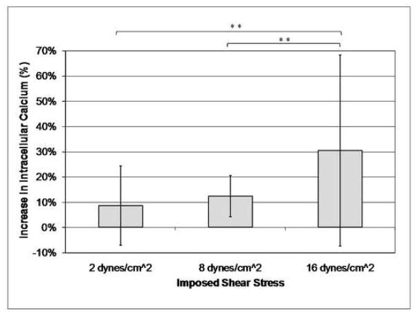
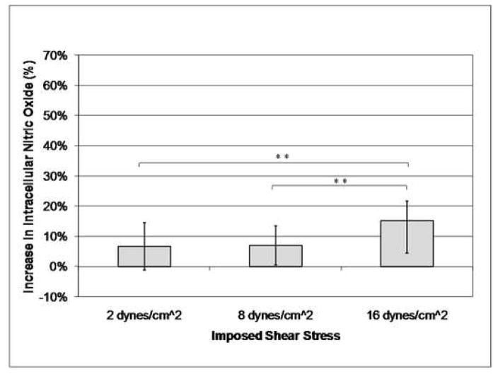
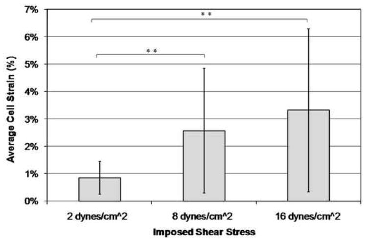
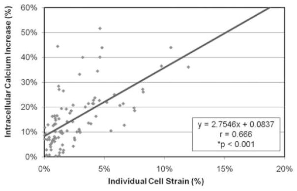
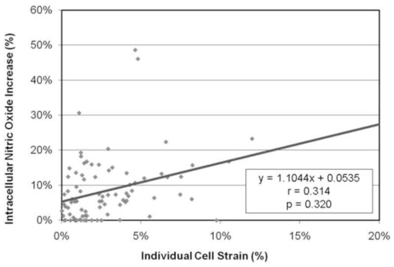
Osteocytes compose 90-95% of all bone cells and are the mechanosensors of bone. In this study, the strain experienced by individual osteocytes resulting from an applied fluid flow shear stress was quantified and correlated to two biological responses measured in real-time within the same individual osteocytes: (1) the upregulation of intracellular calcium and (2) changes in intracellular nitric oxide. Osteocyte-like MLO-Y4 cells were loaded with Fluo-4 AM and DAR-4M and exposed to uniform laminar fluid flow shear stresses of 2, 8, or 16 dyn/cm(2). Intracellular calcium and nitric oxide changes were determined by measuring the difference in fluorescence intensity from the cell's basal level prior to fluid flow and the level immediately following exposure. Individual cell strains were calculated using digital image correlation. MLO-Y4 cells showed a linear increase in cell strain, intracellular calcium concentration, and nitric oxide concentration with an increase in applied fluid flow rate. The increase in intracellular calcium was well correlated to the strain that each cell experienced. This study shows that osteocytes exposed to the same fluid flow experienced a range of individual strains and changes in intracellular calcium and nitric oxide concentrations, and the changes in intracellular calcium were correlated with cell strain. These results are among the first to establish a relationship between the strain experienced by osteocytes in response to fluid flow shear and a biological response at the single cell level. Mechanosensing and chemical signaling in osteocytes has been hypothesized to occur at the single cell level, making it imperative to understand the biological response of the individual cell.
Copyright 2010 Elsevier Ltd. All rights reserved.
Conflict of interest statement
The authors have no conflicts of interest.
Figures
References
-
- Ajubi NE, Klein-Nulend J, Alblas MJ, Burger EH, Nijweide PJ. Signal transduction pathways involved in fluid flow-induced PGE2 production by cultured osteocytes. Am J Physiol. 1999;276(1 Pt 1):E171–8. - PubMed
-
- Aonuma Y, Adachi T, Tanaka M, Hojo M, Takano-Yamamoto T, Kamioka H. Mechanosensitivity of a single osteocyte: Difference in cell process and cell body. Journal of Biomechanical Science and Engineering. 2007;2(S1):S165.
-
- Bacabac RG, Smit TH, Mullender MG, Dijcks SJ, Van Loon JJ, Klein-Nulend J. Nitric oxide production by bone cells is fluid shear stress rate dependent. Biochem Biophys Res Commun. 2004;315(4):823–9. - PubMed
-
- Bacabac RG, Smit TH, Mullender MG, Van Loon JJ, Klein-Nulend J. Initial stress-kick is required for fluid shear stress-induced rate dependent activation of bone cells. Ann Biomed Eng. 2005;33(1):104–10. - PubMed
-
- Bonewald LF. Osteocytes: a proposed multifunctional bone cell. J Musculoskelet Neuronal Interact. 2002;2(3):239–41. - PubMed
Publication types
MeSH terms
Substances
Grants and funding
LinkOut - more resources
Full Text Sources
