

Postnatal somatic cell proliferation and seminiferous tubule maturation in pigs: a non-random event
- PMID: 20189235
- PMCID: PMC4805375
- DOI: 10.1016/j.theriogenology.2009.12.014
Postnatal somatic cell proliferation and seminiferous tubule maturation in pigs: a non-random event
Abstract
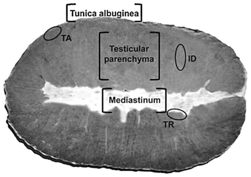
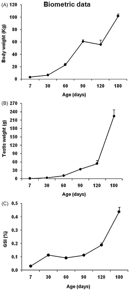
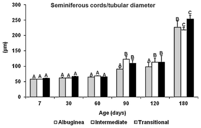
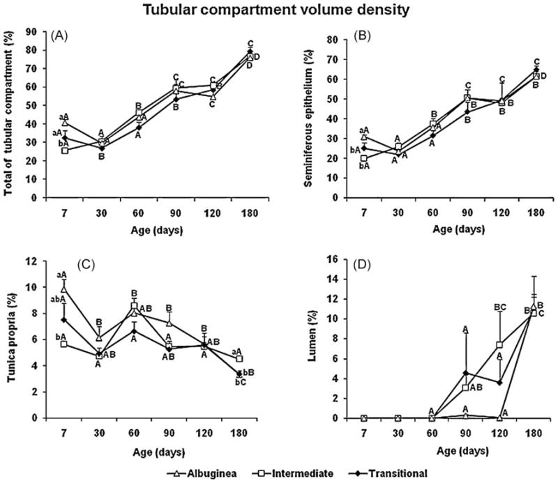
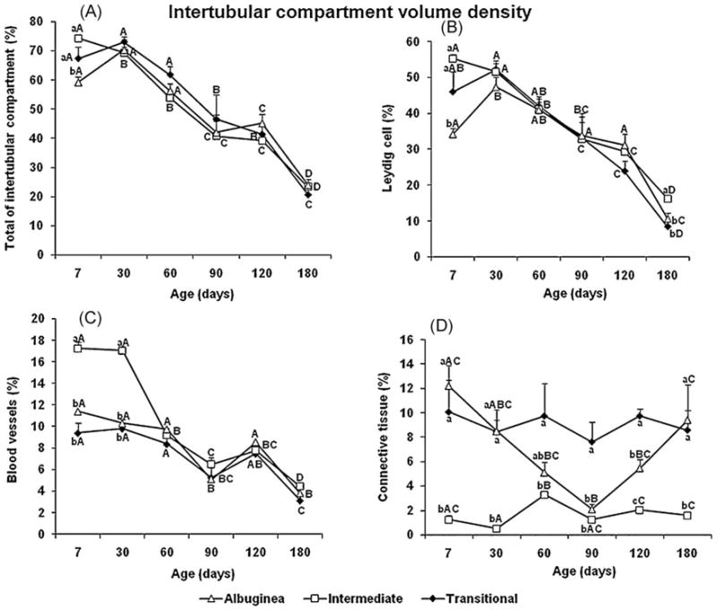
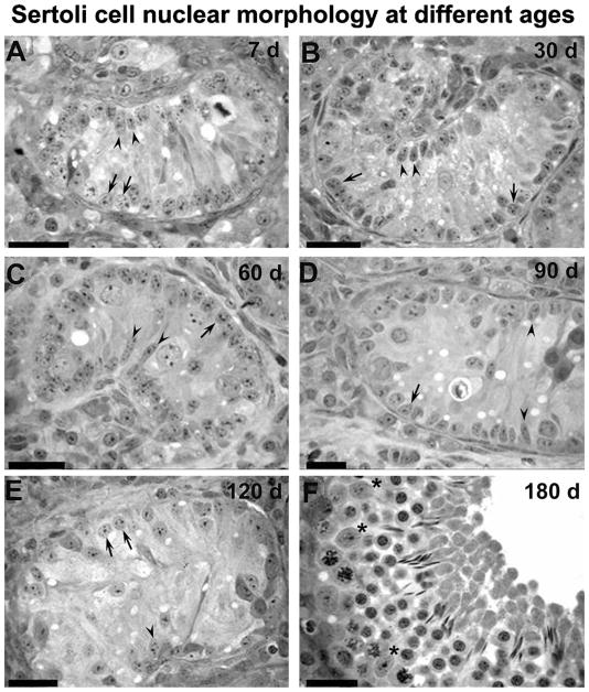
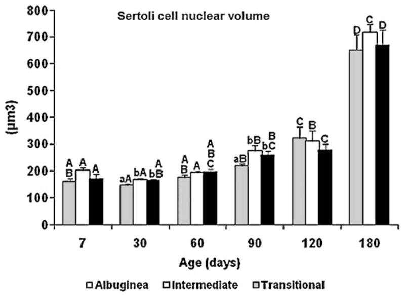
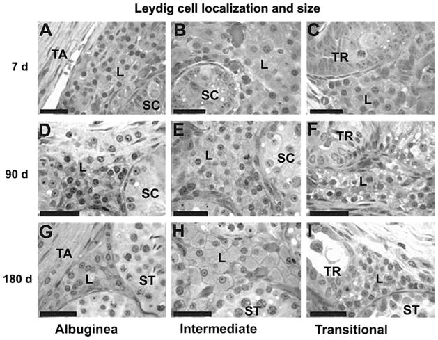
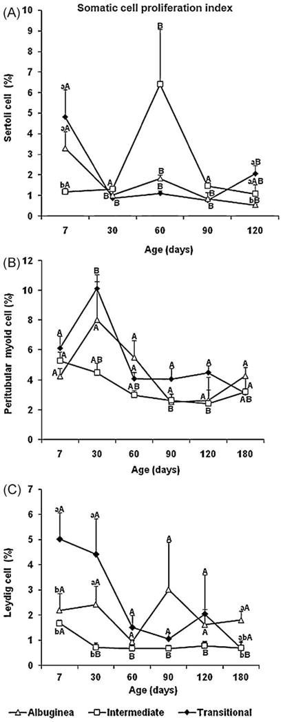
Although seminiferous tubule maturation in horses begins in the central area of the testis, this process is thought to occur randomly throughout the testis in most mammals. Studies in our laboratory revealed that the establishment of spermatogenesis may not be a synchronous event in the testicular parenchyma of pigs. The objectives of the present study were to evaluate the pattern of seminiferous cord/tubule maturation and the morphological and functional characteristics of testicular somatic cells during postnatal development in three regions of the pig testis: a) near the tunica albuginea (TA); b) in the transitional area between the seminiferous tubules and mediastinum (TR); and c) in the intermediate area (ID) between the TA and TR. Based on the diameter of seminiferous cords/tubules, nucleus size of Sertoli cells and fluid secretion, mainly at 90 and 120 d of age, seminiferous tubule maturation was more advanced in the ID and TR. The mitotic activity of Sertoli cells was higher (P<0.05) in the TR than the ID and TA at 7 and 120 d. Except for the mitotic index of the Leydig cells, which was lower (P<0.05) in the ID at 7, 30, and 180 d than in the TA and TR, other Leydig cell ebd points, e.g., individual cell size, nuclear volume, and cytoplasmic volume, were consistently higher (P<0.05) in the ID, suggesting that steroidogenesis was more active in this region during the period investigated. Overall, we inferred that Leydig cells in the ID may play a pivotal role in postnatal testis development in pigs and this type of cell is likely related to asynchronous testicular parenchyma development, with the transitional area providing the primary zone for growth of seminiferous tubules.
Figures

Similar articles
-
Cell proliferation and hormonal changes during postnatal development of the testis in the pig.Biol Reprod. 2000 Dec;63(6):1629-36. doi: 10.1095/biolreprod63.6.1629. Biol Reprod. 2000. PMID: 11090429
-
Development of the seminiferous tubules after neonatal hemicastration in the boar.J Reprod Fertil. 1989 Sep;87(1):1-11. doi: 10.1530/jrf.0.0870001. J Reprod Fertil. 1989. PMID: 2621685
-
Variations in testicular androgen receptors and histology of the lamb testis from birth to puberty.J Reprod Fertil. 1984 Jan;70(1):203-10. doi: 10.1530/jrf.0.0700203. J Reprod Fertil. 1984. PMID: 6694138
-
Ultrastructure of the aging human testis.J Electron Microsc Tech. 1991 Oct;19(2):241-60. doi: 10.1002/jemt.1060190209. J Electron Microsc Tech. 1991. PMID: 1748904 Review.
-
Cell-cell interactions in the control of spermatogenesis as studied using Leydig cell destruction and testosterone replacement.Am J Anat. 1990 May;188(1):3-20. doi: 10.1002/aja.1001880103. Am J Anat. 1990. PMID: 2161173 Review.
Cited by
-
Regulatory Effects of the Kiss1 Gene in the Testis on Puberty and Reproduction in Hezuo and Landrance Boars.Int J Mol Sci. 2023 Nov 24;24(23):16700. doi: 10.3390/ijms242316700. Int J Mol Sci. 2023. PMID: 38069021 Free PMC article.
-
Detection and localization of atypical porcine pestivirus in the testicles of naturally infected, congenital tremor affected piglets.Transbound Emerg Dis. 2022 Jul;69(4):e621-e629. doi: 10.1111/tbed.14355. Epub 2021 Nov 8. Transbound Emerg Dis. 2022. PMID: 34705340 Free PMC article.
-
Precocious puberty in male wild boars: a possible explanation for the dramatic population increase in Germany and Europe.PeerJ. 2021 Jul 20;9:e11798. doi: 10.7717/peerj.11798. eCollection 2021. PeerJ. 2021. PMID: 34322327 Free PMC article.
-
Sertoli cell and spermatogonial development in pigs.J Anim Sci Biotechnol. 2022 Apr 11;13(1):45. doi: 10.1186/s40104-022-00687-2. J Anim Sci Biotechnol. 2022. PMID: 35399096 Free PMC article.
-
Ultrasonographic evaluation of testicular and Pampiniform plexus characteristics in young bulls under different microclimatic conditions in a tropical environment.Sci Rep. 2025 Jul 17;15(1):25940. doi: 10.1038/s41598-025-05522-y. Sci Rep. 2025. PMID: 40676009 Free PMC article.
References
-
- Karl J, Capel B. Sertoli cells of the mouse testis originate from the coelomic epithelium. Dev Biol. 1998;203:323–33. - PubMed
-
- Cool J, Capel B. Mixed signals: development of the testis. Semin Reprod Med. 2009;27:5–13. - PubMed
-
- França LR, Silva VA, Jr, Chiarini-Garcia H, Garcia SK, Debeljuk L. Cell proliferation and hormonal changes during postnatal development of the testis in the pig. Biol Reprod. 2000;63:1629–36. - PubMed
-
- Alvarenga ER, França LR. Effects of different temperatures on testis structure and function, with emphasis on somatic cells, in sexually mature Nile tilapias (Oreochromis niloticus) Biol Reprod. 2009;80:537–44. - PubMed
Publication types
MeSH terms
Grants and funding
LinkOut - more resources
Full Text Sources
