. 2010 Mar 9;20(5):458-63.
doi: 10.1016/j.cub.2010.01.038.

Retinoids regulate a developmental checkpoint for tissue regeneration in Drosophila

Affiliations

Retinoids regulate a developmental checkpoint for tissue regeneration in Drosophila

Adrian Halme et al. Curr Biol. .

Abstract

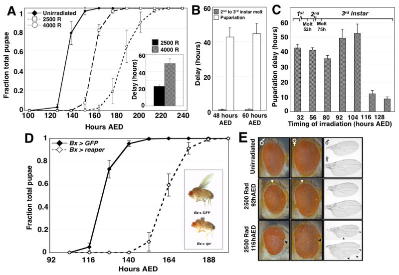
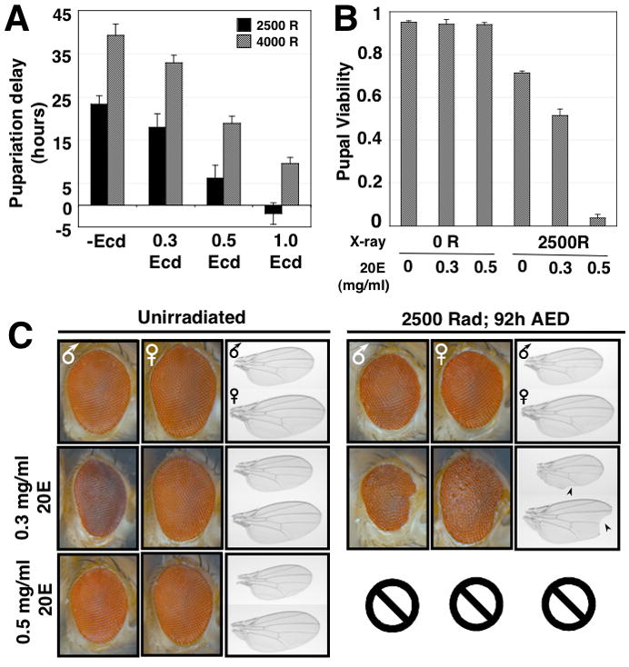
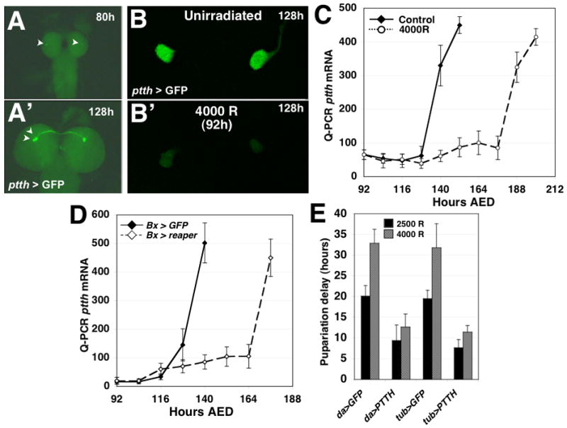
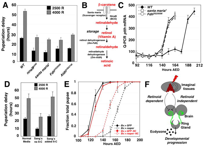
Damage to Drosophila imaginal discs elicits a robust regenerative response from the surviving tissue [1-4]. However, as in other organisms, developmental progression and differentiation can restrict the regenerative capacity of Drosophila tissues. Experiments in Drosophila and other holometabolous insects have demonstrated that either damage to imaginal tissues [5, 6] or transplantation of a damaged imaginal disc [7, 8] delays the onset of metamorphosis. Therefore, in Drosophila there appears to be a mechanism that senses tissue damage and extends the larval phase to coordinate tissue regeneration with the overall developmental program of the organism. However, how such a pathway functions remains unknown. Here we demonstrate that a developmental checkpoint extends larval growth after imaginal disc damage by inhibiting the transcription of the gene encoding PTTH, a neuropeptide that promotes the release of the steroid hormone ecdysone. Using a genetic screen, we identify a previously unsuspected role for retinoid biosynthesis in regulating PTTH expression and delaying development in response to tissue damage. Retinoid signaling plays an important but poorly defined role in several vertebrate regeneration models [9-11]. Our findings demonstrate that retinoid biosynthesis in Drosophila is important for the maintenance of a condition that is permissive for regenerative growth.


Figures

Figure 1
Figure 1
Imaginal tissue damage activates a developmental checkpoint during the third larval instar of Drosophila development. (A) Timing of pupariation for unirradiated larvae, or larvae X-irradiated at 92 hours AED with doses of either 2500 rads or 4000 rads. Developmental timing is represented as both the fraction of total larvae pupariated and in the inset graph as the difference between the median timing to pupariation of irradiated and unirradiated populations of larvae (delay). In all developmental timing experiments described in this paper, n=3 independent populations were assayed for each condition in each genetic background unless otherwise noted. Error bars in all figures represent the standard error for each data point. (B) Duration of delay for both second to third instar molting (gray bars) and pupariation (solid white bars) among larvae irradiated with 4000 rad at either 48 hours AED, during the first larval instar, or at 60 hours AED, during the second larval instar. (C) Pupariation delay for larvae irradiated with 4000 rad at progressively later times throughout larval development. (D) Timing of pupariation for Bx > rpr and Bx > GFP larvae. Bx > rpr larvae exhibit a substantial delay in pupariation timing. (Inset) adult flies expressing either UAS-GFP or UAS-reaper under the control of Bx-GAL4. Eclosed Bx > rpr flies have ablated wings, but exhibit no other developmental or obvious behavioral abnormalities and are fertile. (E) Eyes (shown with posterior to the left) and wings (proximal to the left) from male and female adults irradiated as larvae either before (92 hours AED) or after (116 hours AED) the developmental checkpoint. The boundary of posterior ommatidial disorganization is shown by the white triangles. Anterior notching of eyes is marked with black triangles. Wing notching is marked with arrowheads.
Figure 2
Figure 2
Tissue regeneration and pupal viability in X-irradiated larvae is compromised by 20E addition. (A) Developmental delay of larvae exposed to 2500 or 4000 rad X-irradiation, and fed 0.3, 0.5 or 1.0 mg/ml 20E dissolved in 100% ethanol, or an equivalent volume of ethanol as a control. (B) Pupal viability (as determined by the initiation of adult eclosion from the pupal case) of either unirradiated larvae or larvae irradiated at 92 hours AED, that were either fed different concentrations of 20E (0.3mg/ml and 0.5 mg/ml) in Drosophila molasses-cornmeal media or an equivalent volume of ethanol in molasses-cornmeal media as a negative control. The viability for n=3 independent populations was assayed for each condition (C) Eyes and wings from male and female adult flies irradiated as larvae at 92 hours AED, then either fed 0.3, 0.5 or 1.0 mg/ml 20E or an equivalent volume of ethanol in their normal food. Wing notching is shown with arrowheads. Two larvae irradiated and fed 0.5mg/ml 20E eclosed (Fig. 2B), however one adult died before completing eclosion, and the second adult died immediately in the food. Therefore we were unable to effectively assess the wing and eye phenotypes in these animals.
Figure 3
Figure 3
Irradiation-induced tissue damage inhibits PTTH expression and endocrine signaling. (A) Larvae carrying PTTH-GAL4 and UAS-GFP transgenes can be used to visualize PTTH gene activity in PTTH-expressing neurons in the larval brain. The previously described [18] upregulation of PTTH gene activity during developmental progression can be visualized as an increase in GFP fluorescence in the neuron cell bodies (arrowheads) of the PTTH-expressing neurons (A: PTTH > GFP expression in a larval brain fixed at 80 hours AED; A′: PTTH > GFP expression at 128 hours AED). (B) PTTH > GFP expression at 128 hours AED in the brain of an unirradiated larvae or a larva that was X-irradiated at 92 hours AED with 4000 rad. (C) Measurement of ptth transcript levels in unirradiated and X-irradiated (4000 rad) larvae. Larvae were collected every 12 hours after irradiation until the animals had entered pupal phase of development. ptth gene transcript levels were assayed with quantitative RT-PCR with probes targeting the ptth transcript. (D) ptth transcriptional activity in Bx > rpr and Bx > GFP larvae. Bx > rpr larvae exhibit a delayed upregulation of ptth transcript that is consistent with their delayed developmental timing. ptth transcript was detected using quantitative RT-PCR. (E) Ectopic expression of ptth attenuates irradiation-induced developmental delays. Larvae carrying either the daughterless-GAL4 or the tubulin-GAL4 expression transgenes were crossed to either lines carrying UAS-GFP or UAS-ptth. In the progeny of these crosses we assayed the effects of da > PTTH and tub > PTTH expression on the pupariation delay of larvae irradiated at 92 hours AED. Larvae expressing da > GFP or tub > GFP were assayed as controls for developmental delay. n=4 populations were assayed for each set of conditions in each genetic background.
Figure 4
Figure 4
Retinoid metabolism regulates checkpoint-induced delay in irradiated larvae. (A) Pupariation delay in wild-type larvae or homozygous mutant larvae carrying mutations in genes required for retinoid metabolism. n=3 independent populations were assayed for all timing experiments with all mutants unless otherwise noted. p < 0.01 for each pairwise comparison of wild-type and mutant pupariation delay for all four mutants irradiated at 2500 and 4000 rad, as calculated by two-tailed Student’s t-test. (B) A schematic illustrating retinoid metabolic pathways and the gene products required for each step of retinoid metabolism. The genes responsible for retinol dehydrogenase and retinaldehyde dehydrogenase activities in Drosophila have not been identified. Sequence comparisons suggest that the most similar Drosophila gene to vertebrate retinol dehydrogenase is the Drosophila Formaldehyde dehydrogenase (Fdh) gene; the most similar Drosophila gene to vertebrate retinaldehyde dehydrogenase is the Aldehyde dehydrogenase (Aldh) gene of Drosophila. (C) ptth transcript levels in X-irradiated (4000 R), wild type, FdhDG29306 and santa maria1 larvae. ptth transcript was detected using quantitative RT-PCR. (D) Pupariation delay in irradiated wild-type larvae raised either on normal molasses-cornmeal food, or Sang’s defined medium that is either missing (−A) or supplemented with (+A) β-carotene. p < 0.01 in a pairwise comparison of delay in Normal vs. Sang’s (−A) medium at both at 2500 and 4000 rad. The timing of n=3 independent populations were assayed for each condition. p < 0.01 in a pairwise comparison of delay in Sang’s (−A) vs. Sang’s (+A) medium at both at 2500 and 4000 rad; both p-values were calculated by two-tailed Student’s t-test. (E) Developmental timing of Bx > GFP and Bx > rpr larvae raised on carotenoid-deficient Sang’s defined medium (red) or Sang’s defined medium supplemented with 1.25 g/L β-carotene (black). Bx > rpr larvae grown on carotenoid-deficient Sang’s medium exhibit a shorter larval duration than siblings grown on β-carotene supplemented medium. The timing of n=4 independent populations were assayed for each genetic background under each condition. (F) A model for the developmental checkpoint for tissue repair. Damage to imaginal discs is capable of regulating the expression of the ptth gene in the PTTH-expressing neurons via both a retinoid-dependent pathway, defined by the retinoid biosynthesis mutant phenotypes described in this work, and a second, retinoid-independent pathway.

References

    1. Bryant P. Regeneration and duplication in imaginal discs. Ciba Foundation symposium.1975. - PubMed
    1. Bryant P. Pattern formation in the imaginal wing disc of Drosophila melanogaster: fate map, regeneration and duplication. J Exp Zool. 1975;193:49–77. - PubMed
    1. Schubiger G. Regeneration, duplication and transdetermination in fragments of the leg disc of Drosophila melanogaster. Dev Biol. 1971;26:277–295. - PubMed
    1. Smith-Bolton RK, Worley MI, Kanda H, Hariharan IK. Regenerative growth in Drosophila imaginal discs is regulated by Wingless and Myc. Dev Cell. 2009;16:797–809. - PMC - PubMed
    1. Hussey R, Thompson W, Calhoun E. The influence of x-rays on the development of drosophila larvae. Science. 1927;66:65–66. - PubMed

Publication types