. 2010 Feb 26;32(2):175-86.
doi: 10.1016/j.immuni.2010.02.004.

Synergistic signals for natural cytotoxicity are required to overcome inhibition by c-Cbl ubiquitin ligase

Affiliations

Synergistic signals for natural cytotoxicity are required to overcome inhibition by c-Cbl ubiquitin ligase

Hun Sik Kim et al. Immunity. .

Abstract

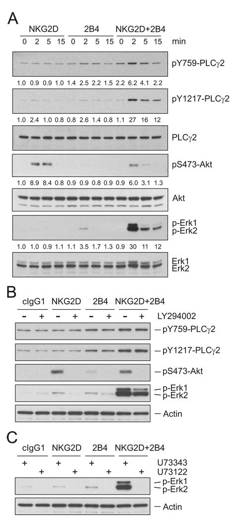
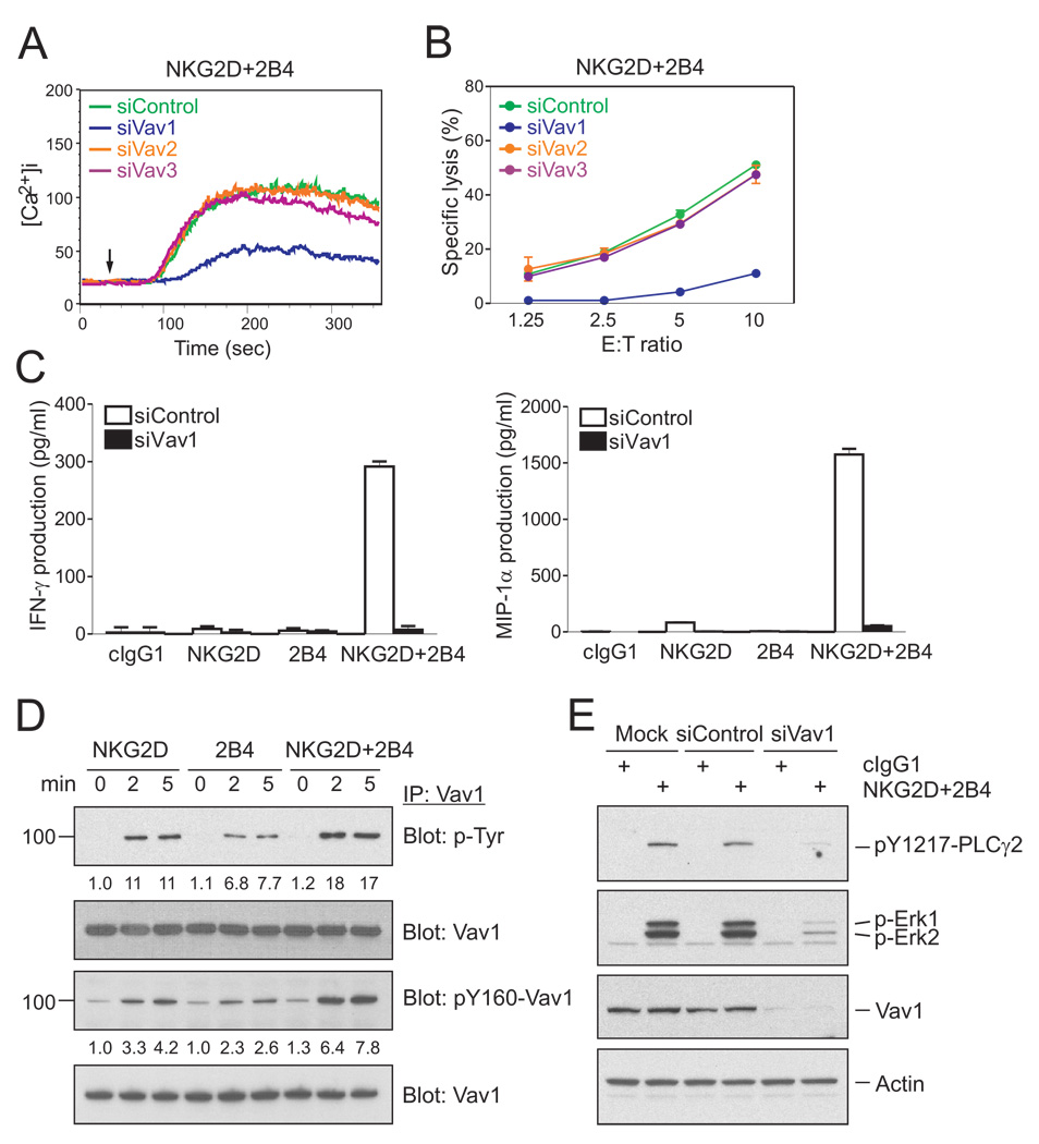
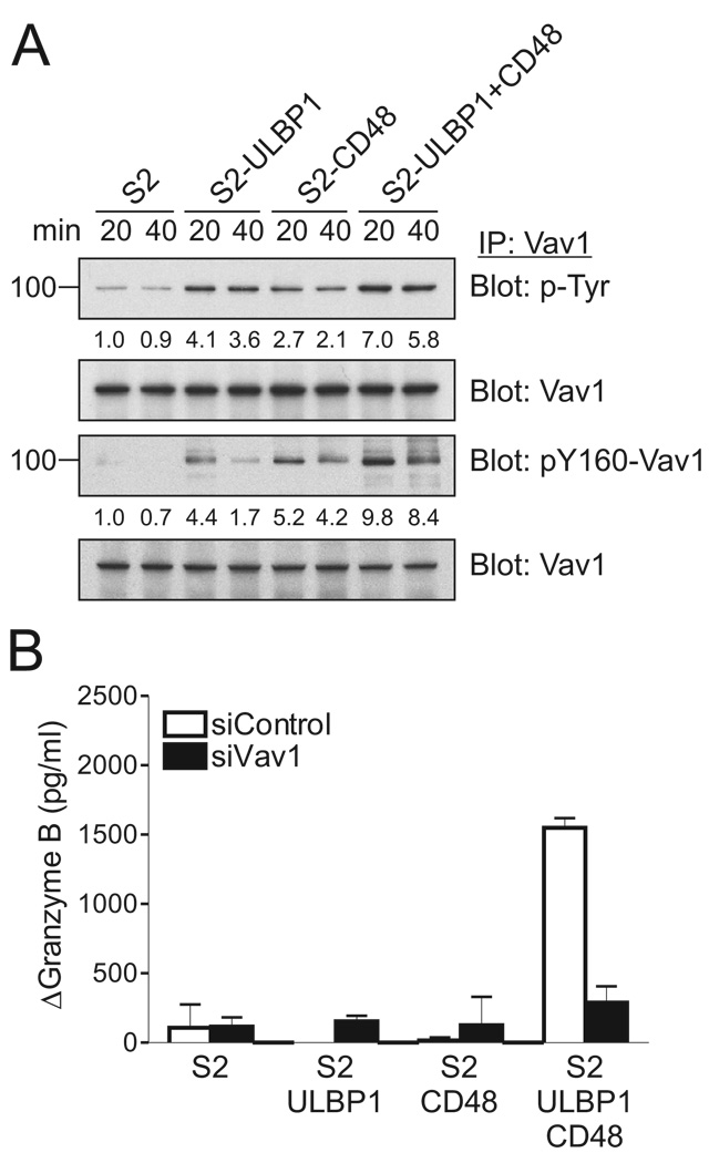
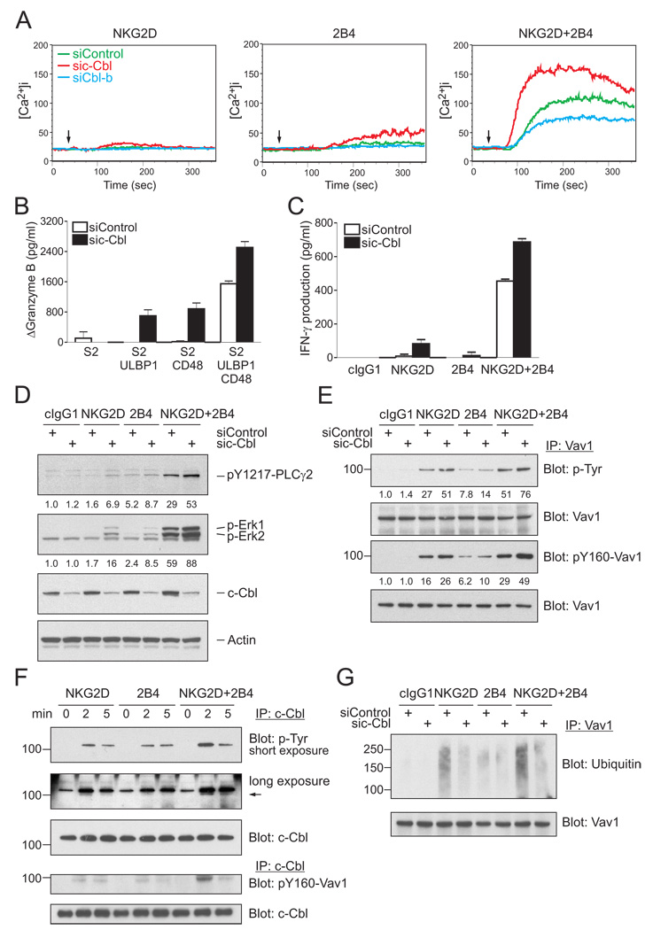
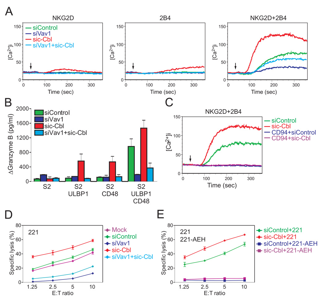
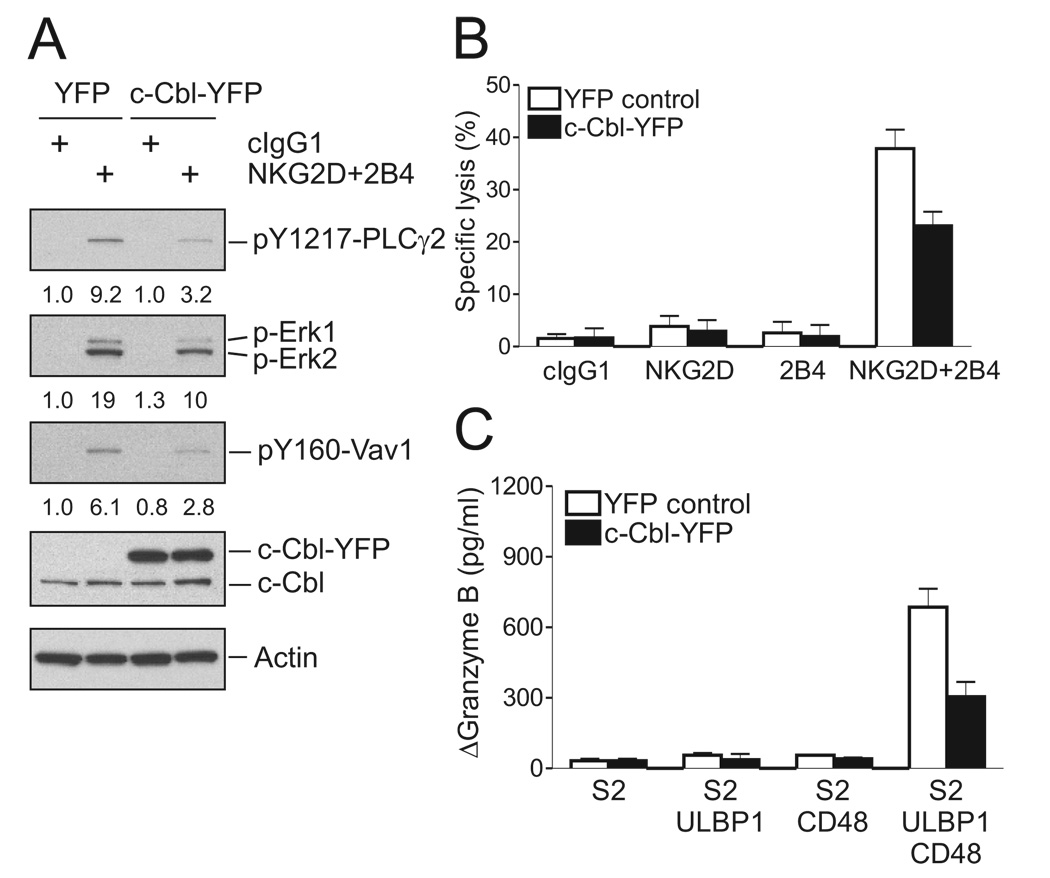
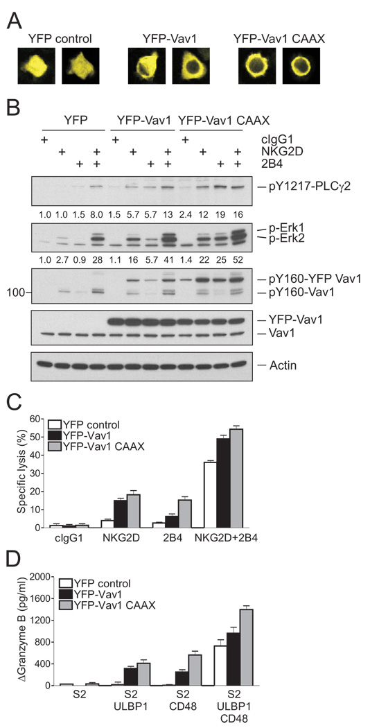
Natural killer (NK) cell cytotoxicity toward target cells depends on synergistic coactivation by NK cell receptors such as NKG2D and 2B4. How synergy occurs is not known. Synergistic phosphorylation of phospholipase PLC-gamma2, Ca(2+) mobilization, and degranulation triggered by NKG2D and 2B4 coengagement were blocked by Vav1 siRNA knockdown, but enhanced by knockdown of c-Cbl. c-Cbl inhibited Vav1-dependent signals, given that c-Cbl knockdown did not rescue the Vav1 defect. Moreover, c-Cbl knockdown and Vav1 overexpression each circumvented the necessity for synergy because NKG2D or 2B4 alone became sufficient for activation. Thus, synergy requires not strict complementation but, rather, strong Vav1 signals to overcome inhibition by c-Cbl. Inhibition of NK cell cytotoxicity by CD94-NKG2A binding to HLA-E on target cells was dominant over synergistic activation, even after c-Cbl knockdown. Therefore, NK cell activation by synergizing receptors is regulated at the level of Vav1 by a hierarchy of inhibitory mechanisms.


Figures

Figure 1
Figure 1. Synergistic Phosphorylation of PLC-γ2 by NKG2D and 2B4 Coengagement
(A) Freshly isolated, resting NK cells were preincubated with mAbs specific for NKG2D and/or 2B4 on ice for 30 min and stimulated by cross-linking with secondary goat F(ab′)2 anti-mouse IgG at 37°C for the indicated time. Lysates were resolved on a 4 to 12% SDS-PAGE, transferred to a polyvinylidene difluoride (PVDF) membrane, and immunoblotted with Abs to phospho-PLC-γ2 at tyrosine 759 (pY759) or at tyrosine 1217 (pY1217), phospho-Akt at serine 473 (pS473) or phospho-Erk1 and 2. After stripping, the blots were re-probed with Abs to PLC-γ2, Akt, and Erk1 and Erk2. The normalized intensities of the phosphorylated PLC-γ2, Akt, and Erk1 and 2 relative to their total forms were quantified with ImageJ software and are presented for each condition. (B) Resting NK cells were preincubated with isotype control mAb or mAbs specific for NKG2D and/or 2B4 in the absence or presence of the PI3K inhibitor LY294002 at 20 µM for 30 min. After receptor cross-linking for 2 min in the presence of the inhibitor, cells were lysed and the lysates were immunoblotted using Abs to phospho-PLC-γ2 at tyrosine 759 (pY759) or at tyrosine 1217 (pY1217), phospho-Akt at serine 473 (pS473) or phospho-Erk1 and 2. The blot was stripped and re-probed for actin to monitor sample loading. (C) Resting NK cells were preincubated with isotype control mAb or mAbs specific for NKG2D and/or 2B4 in the presence of a PLC-γ inhibitor (U73122; 5 µM) or its inactive analog (U73343; 5 µM) for 30 min. After receptor cross-linking for 2 min in the presence of inhibitors, cells were lysed and the lysates were immunoblotted with Abs to phospho-Erk1 and 2. The blot was stripped and re-probed for actin. Data are representative of at least four independent experiments.
Figure 2
Figure 2. Vav1 Is Required for NKG2D and 2B4 Synergy
(A) NKL cells were transfected with 300 pmoles of control siRNA or siRNA specific for Vav1, Vav2, or Vav3. After 24 hr, the cells were rested in 5% FBS-containing medium in the absence of IL-2 for another 24 hr, loaded with Fluo-4 at 30°C for 30 min, resuspended in HBSS containing 1% FBS, and preincubated with mAbs for NKG2D and 2B4. Cells were pre-warmed at 37°C and analyzed by flow cytometry. After 30 s, goat F(ab′)2 anti-mouse IgG was added to cross-link the receptors. Changes in fluorescence are shown as a function of time. (B) Redirected lysis of P815 cells by rested NKL cells transfected with the indicated siRNAs at the indicated effector to target cell ratios (E:T). Cytotoxicity against P815 cells preincubated with mAbs specific for NKG2D and 2B4 was determined after 2 hr with the Europium assay. Error bars represent the SD. (C) Cytokine release assays with rested NKL cells after transfection with control siRNA or Vav1-specific siRNA and stimulation with beads coated with isotype control mAb (cIgG1) or mAbs specific for NKG2D and/or 2B4. After incubation for 16 hr, IFN-γ and MIP-1α in the supernatants were measured by ELISA. Values represent mean ± SD. (D) Rested NKL cells were treated as in Figure 1A to stimulate NKG2D and/or 2B4 at 37°C for the indicated time. Lysates were immunoprecipitated with Ab to Vav1, immunoblotted with anti-p-Tyr mAb or anti-pY160-Vav1 Ab, and re-probed for Vav1. The normalized intensities of the phosphorylated Vav1 at tyrosines or at tyrosine 160 relative to total Vav1 are presented. (E) Rested NKL cells that were mock-transfected or transfected with control siRNA or Vav1-specific siRNA were stimulated with isotype control mAb (cIgG1) or mAbs specific for NKG2D and 2B4. After receptor cross-linking for 2 min, lysates were immunoblotted for pY1217-PLC-γ2, p-Erk1 and 2, or Vav1. After stripping, the blots were re-probed for actin. Data are representative of at least four independent experiments.
Figure 3
Figure 3. Coactivation by NKG2D and 2B4 Ligands Is Vav1-Dependent
(A) Rested NKL cells were mixed with S2, S2-ULBP1, S2-CD48, or S2-ULBP1+CD48 insect cells, as indicated. After incubation for 20 or 40 min, Vav1 was immunoprecipitated from lysates and immunoblotted for p-Tyr and pY160-Vav1. Blots were re-probed for Vav1. The normalized intensities of the phosphorylated Vav1 at tyrosines or at tyrosine 160 relative to total Vav1 are presented. (B) Granzyme B release assay with rested NKL cells that were transfected with control siRNA or Vav1-specific siRNA and incubated with S2 cells or S2 cells expressing ULBP1 and/or CD48, as indicated. After incubation for 2 hr, granzyme B in the supernatants was measured by ELISA. The increase of granzyme B after incubation with target cells relative to granzyme B upon incubation of NKL cells alone (ΔGranzyme B) is presented. Values represent mean ± SD. Data are representative of at least three independent experiments.
Figure 4
Figure 4. c-Cbl Limits NK Cell Activation and Imposes a Requirement for Synergy
(A) Calcium flux in rested NKL cells transfected with either control siRNA or siRNA specific for c-Cbl or Cbl-b and stimulated with NKG2D and 2B4 as in Figure 2A. (B) Granzyme B released by rested NKL cells transfected with control siRNA or c-Cbl-specific siRNA and incubated with S2 cells or S2 cells expressing ULBP1, CD48, or both, as in Figure 3B. Values (ΔGranzyme B) represent mean ± SD. (C) IFN-γ released by rested NKL cells transfected with control siRNA or c-Cbl-specific siRNA and stimulated with beads coated with isotype control mAb (cIgG1) or mAbs to NKG2D and 2B4 as in Figure 2C. Values represent mean ± SD. (D) Rested NKL cells transfected with control siRNA or c-Cbl-specific siRNA were stimulated with isotype control mAb (cIgG1) or mAbs to NKG2D and 2B4. After receptor cross-linking for 2 min, lysates were immunoblotted for pY1217-PLC-γ2, p-Erk1 and 2, and c-Cbl. Blots were re-probed for actin. The normalized intensities of the phosphorylated PLC-γ2 and Erk1 and 2 relative to actin are presented. (E) Rested NKL cells were treated as in Figure 1A to stimulate NKG2D and 2B4 at 37°C for the indicated time. c-Cbl was immunoprecipitated from lysates and probed with a mAb to p-Tyr (top) and an Ab to pY160-Vav1 (bottom). The arrow points to a distinct tyrosine-phosphorylated protein associated with c-Cbl, which was visible after a long exposure. The blots were stripped and re-probed for c-Cbl. (F and G) Rested NKL cells transfected with control siRNA or c-Cbl-specific siRNA were stimulated with isotype control mAb (cIgG1) or mAbs to NKG2D, 2B4, or both. After receptor cross-linking for 2 min, Vav1 was immunoprecipitated from lysates and probed with Abs to p-Tyr or pY160-Vav1 (F), or an Ab to ubiquitin (G). Blots were re-probed for Vav1. The normalized intensities of the phosphorylated Vav1 at tyrosines or at tyrosine 160 relative to total Vav1 are presented. Data are representative of at least four independent experiments.
Figure 5
Figure 5. Signals Inhibited by c-Cbl Are Vav1-Dependent
(A) Calcium flux in rested NKL cells transfected with either control siRNA or siRNAs specific for Vav1 and for c-Cbl. NKL cells were stimulated with Abs to NKG2D and 2B4 as in Figure 2A. (B) Granzyme B released by rested NKL cells transfected with control siRNA or siRNAs specific for Vav1 and for c-Cbl and incubated with S2 cells or S2 cells expressing ULBP1, CD48, or both, as in Figure 3B. Values (ΔGranzyme B) represent mean ± SD. (C) Calcium flux in rested NKL cells transfected with either control siRNA or siRNA specific for c-Cbl and stimulated with Abs to NKG2D and 2B4 combined with or without a mAb to CD94, as in Figure 2A. (D) Lysis of 221 cells by rested NKL cells either mock-transfected or transfected with control siRNA or siRNAs specific for Vav1 and for c-Cbl. (E) Lysis of 221 or 221-AEH cells by rested NKL cells transfected with control siRNA or siRNA specific for c-Cbl. Error bars represent the SD. Data are representative of at least three independent experiments.
Figure 6
Figure 6. c-Cbl Overexpression Inhibits NKG2D and 2B4 Synergy
NKL cells were transfected with 8 µg of YFP vector control or c-Cbl-YFP plasmid. After 24 hr, cells were sorted for YFP+ cells. (A) Rested NKL cells transfected with the indicated plasmid were stimulated with isotype control mAb (cIgG1) or mAbs to NKG2D and 2B4. After receptor cross-linking for 2 min, lysates were immunoblotted for pY1217-PLC-γ2, p-Erk1 and 2, and pY160-Vav1. Blots were re-probed for c-Cbl and actin. The normalized intensities of the phosphorylated PLC-γ2, Erk1 and 2, and Vav1 at tyrosine 160 relative to actin are presented. (B) Redirected lysis of P815 cells by rested NKL cells transfected with the indicated plasmid at an effector to target cell ratio of 10. NKL cells were stimulated with mAbs to NKG2D, 2B4, or both. Error bars represent the SD. (C) Granzyme B released by rested NKL cells transfected with the indicated plasmid and incubated with S2 cells or S2 cells expressing ULBP1, CD48, or both, as in Figure 3B. Values (ΔGranzyme B) represent mean ± SD. Data are representative of at least three independent experiments.
Figure 7
Figure 7. Increased Vav1 Expression Overcomes Inhibition by c-Cbl
NKL cells were transfected with 8 µg of YFP vector control, YFP-Vav1, or YFP-Vav1 CAAX plasmid. After 24 hr, cells were sorted for YFP+ cells. (A) Representative confocal images of NKL cells transfected with the indicated plasmid. (B) Rested NKL cells transfected with the indicated plasmid were stimulated with isotype control mAb (cIgG1) or mAbs to NKG2D, 2B4, or both. After receptor cross-linking for 2 min, lysates were immunoblotted for pY1217-PLC-γ2, p-Erk1 and 2, and pY160-Vav1. Blots were re-probed for Vav1 and actin. The normalized intensities of the phosphorylated PLC-γ2 and Erk1 and 2 relative to actin are presented. (C) Redirected lysis of P815 cells by rested NKL cells transfected with the indicated plasmid at an effector to target cell ratio of 10. NKL cells were stimulated with mAbs to NKG2D, 2B4, or both. Error bars represent the SD. (D) Granzyme B released by rested NKL cells transfected with the indicated plasmid and incubated with S2 cells or S2 cells expressing ULBP1, CD48, or both, as in Figure 3B. Values (ΔGranzyme B) represent mean ± SD. Data are representative of at least three independent experiments.

Similar articles

Cited by

References

    1. Acuto O, Michel F. CD28-mediated co-stimulation: a quantitative support for TCR signalling. Nat Rev Immunol. 2003;3:939–951. - PubMed
    1. Anfossi N, Andre P, Guia S, Falk CS, Roetynck S, Stewart CA, Breso V, Frassati C, Reviron D, Middleton D, et al. Human NK cell education by inhibitory receptors for MHC class I. Immunity. 2006;25:331–342. - PubMed
    1. Anikeeva N, Somersalo K, Sims TN, Thomas VK, Dustin ML, Sykulev Y. Distinct role of lymphocyte function-associated antigen-1 in mediating effective cytolytic activity by cytotoxic T lymphocytes. Proc Natl Acad Sci U S A. 2005;102:6437–6442. - PMC - PubMed
    1. Ardouin L, Bracke M, Mathiot A, Pagakis SN, Norton T, Hogg N, Tybulewicz VLJ. Vav1 transduces TCR signals required for LFA-1 function and cell polarization at the immunological synapse. Eur J Immunol. 2003;33:790–797. - PubMed
    1. Bachmaier K, Krawczyk C, Kozieradzki I, Kong YY, Sasaki T, Oliveira-dos-Santos A, Mariathasan S, Bouchard D, Wakeham A, Itie A, et al. Negative regulation of lymphocyte activation and autoimmunity by the molecular adaptor Cbl-b. Nature. 2000;403:211–216. - PubMed

Publication types

MeSH terms

Substances