

Immunization of cattle with recombinant Newcastle disease virus expressing bovine herpesvirus-1 (BHV-1) glycoprotein D induces mucosal and serum antibody responses and provides partial protection against BHV-1
- PMID: 20189484
- PMCID: PMC3428038
- DOI: 10.1016/j.vaccine.2010.02.051
Immunization of cattle with recombinant Newcastle disease virus expressing bovine herpesvirus-1 (BHV-1) glycoprotein D induces mucosal and serum antibody responses and provides partial protection against BHV-1
Abstract
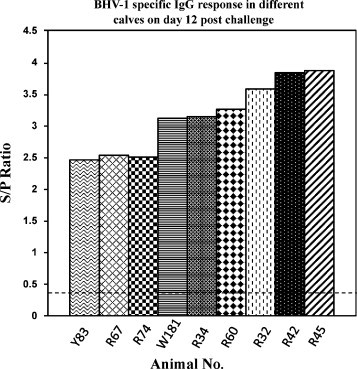
Bovine herpesvirus-1 (BHV-1) is a major cause of respiratory tract diseases in cattle. Vaccination of cattle against BHV-1 is a high priority. A major concern of currently modified live BHV-1 vaccines is their ability to cause latent infection and subsequent reactivation resulting in many outbreaks. Thus, there is a need for alternative strategies. We generated two recombinant Newcastle disease viruses (NDVs) expressing the glycoprotein D (gD) of BHV-1 from an added gene. One recombinant, rLaSota/gDFL, expressed gD without any modification. The other recombinant, rLaSota/gDF, expressed a chimeric gD in which the ectodomain of gD was fused with the transmembrane domain and cytoplasmic tail of the NDV fusion F glycoprotein. Remarkably, the native gD expressed by rLaSota/gDFL virus was incorporated into the NDV virion 2.5-fold more efficiently than the native NDV proteins, whereas the chimeric gD was not detectably incorporated even though it was abundantly expressed on the infected cell surface. The expression of gD did not increase the virulence of the rNDV vectors in chickens. A single intranasal and intratracheal inoculation of calves with either recombinant NDV elicited mucosal and systemic antibodies specific to BHV-1, with the responses to rLaSota/gDFL being higher than those to rLaSota/gDF. Following challenge with BHV-1, calves immunized with the recombinant NDVs had lower titers and earlier clearance of challenge virus compared to the empty vector control, and reduced disease was observed with rLaSota/gDFL. Following challenge, the titers of serum antibodies specific to BHV-1 were higher in the animals immunized with the rNDV vaccines compared to the rNDV parent virus, indicating that the vaccines primed for secondary responses. Our data suggest that NDV can be used as a vaccine vector in bovines and that BHV-1 gD may be useful in mucosal vaccine against BHV-1 infection, but might require augmentation by a second dose or the inclusion of additional BHV-1 antigens.
Copyright 2010 Elsevier Ltd. All rights reserved.
Figures
Similar articles
-
A recombinant Newcastle disease virus (NDV) expressing infectious laryngotracheitis virus (ILTV) surface glycoprotein D protects against highly virulent ILTV and NDV challenges in chickens.Vaccine. 2014 Jun 12;32(28):3555-63. doi: 10.1016/j.vaccine.2014.04.068. Epub 2014 Apr 30. Vaccine. 2014. PMID: 24793943
-
The immunogenicity and efficacy of replication-defective and replication-competent bovine adenovirus-3 expressing bovine herpesvirus-1 glycoprotein gD in cattle.Vet Immunol Immunopathol. 2000 Oct 31;76(3-4):257-68. doi: 10.1016/s0165-2427(00)00217-8. Vet Immunol Immunopathol. 2000. PMID: 11044558
-
Mucosal immunization of calves with recombinant bovine adenovirus-3: induction of protective immunity to bovine herpesvirus-1.J Gen Virol. 1999 May;80 ( Pt 5):1263-1269. doi: 10.1099/0022-1317-80-5-1263. J Gen Virol. 1999. PMID: 10355773
-
A review of the biology of bovine herpesvirus type 1 (BHV-1), its role as a cofactor in the bovine respiratory disease complex and development of improved vaccines.Anim Health Res Rev. 2007 Dec;8(2):187-205. doi: 10.1017/S146625230700134X. Anim Health Res Rev. 2007. PMID: 18218160 Review.
-
DNA immunization with bovine herpesvirus-1 genes.Ann N Y Acad Sci. 1995 Nov 27;772:47-63. doi: 10.1111/j.1749-6632.1995.tb44731.x. Ann N Y Acad Sci. 1995. PMID: 8546413 Review. No abstract available.
Cited by
-
Newcastle disease virus: current status and our understanding.Virus Res. 2014 May 12;184:71-81. doi: 10.1016/j.virusres.2014.02.016. Epub 2014 Mar 1. Virus Res. 2014. PMID: 24589707 Free PMC article. Review.
-
Immune response after oral immunization of goats and foxes with an NDV vectored rabies vaccine candidate.PLoS Negl Trop Dis. 2024 Feb 26;18(2):e0011639. doi: 10.1371/journal.pntd.0011639. eCollection 2024 Feb. PLoS Negl Trop Dis. 2024. PMID: 38408125 Free PMC article.
-
A Novel Recombinant Newcastle Disease Virus Vectored DIVA Vaccine against Peste des Petits Ruminants in Goats.Vaccines (Basel). 2020 Apr 28;8(2):205. doi: 10.3390/vaccines8020205. Vaccines (Basel). 2020. PMID: 32354145 Free PMC article.
-
A Scalable Topical Vectored Vaccine Candidate against SARS-CoV-2.Vaccines (Basel). 2020 Aug 24;8(3):472. doi: 10.3390/vaccines8030472. Vaccines (Basel). 2020. PMID: 32846910 Free PMC article.
-
Characterization of a recombinant Newcastle disease virus expressing the glycoprotein of bovine ephemeral fever virus.Arch Virol. 2017 Feb;162(2):359-367. doi: 10.1007/s00705-016-3078-2. Epub 2016 Oct 18. Arch Virol. 2017. PMID: 27757685 Free PMC article.
References
-
- van Drunen Littel-van den Hurk S., Parker M.D., Massie B., van den Hurk J.V., Harland R., Babiuk L.A. Protection of cattle from BHV-1 infection by immunization with recombinant glycoprotein gIV. Vaccine. 1993;11(1):25–35. - PubMed
-
- Babiuk L.A., L’Italien J., van Drunen Littel-van den Hurk S., Zamb T., Lawman J.P., Hughes G. Protection of cattle from bovine herpesvirus type I (BHV-1) infection by immunization with individual viral glycoproteins. Virology. 1987;159(July (1)):57–66. - PubMed
Publication types
MeSH terms
Substances
Grants and funding
LinkOut - more resources
Full Text Sources
Other Literature Sources
