Comparative Study
. 2011 Dec;32(12):2198-210.
doi: 10.1016/j.neurobiolaging.2010.01.009. Epub 2010 Mar 1.

Age-related changes in Arc transcription and DNA methylation within the hippocampus

Affiliations
Comparative Study

Age-related changes in Arc transcription and DNA methylation within the hippocampus

M R Penner et al. Neurobiol Aging. 2011 Dec.

Abstract

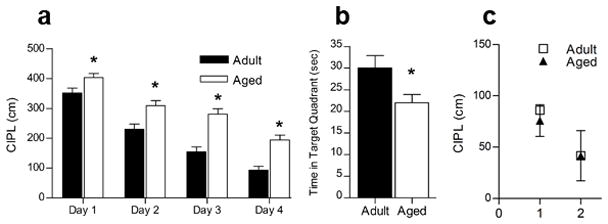
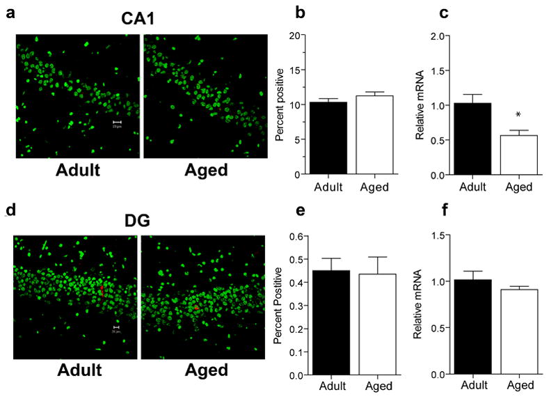
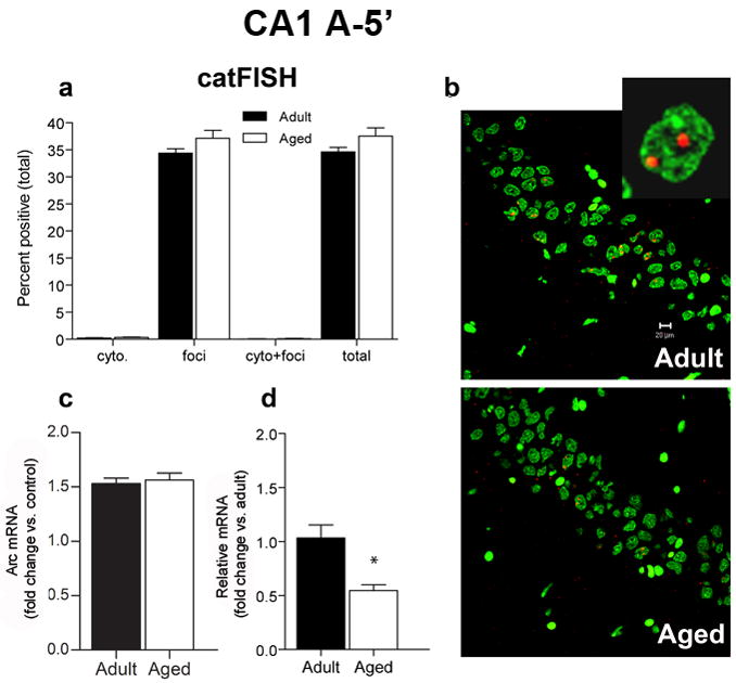
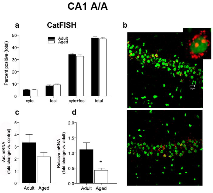
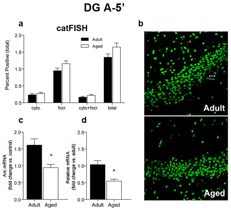
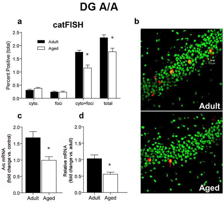
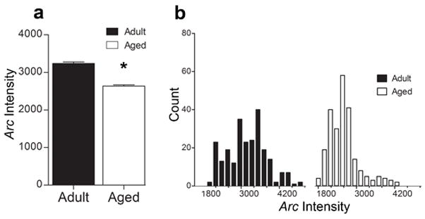
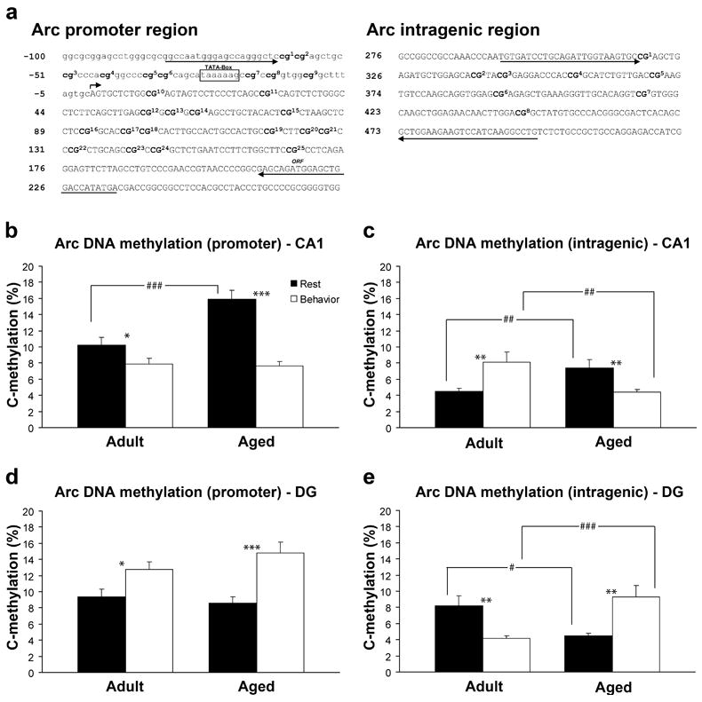
The transcription of genes that support memory processes are likely to be impacted by the normal aging process. Because Arc is necessary for memory consolidation and enduring synaptic plasticity, we examined Arc transcription within the aged hippocampus. Here, we report that Arc transcription is reduced within the aged hippocampus compared to the adult hippocampus during both "off line" periods of rest, and following spatial behavior. This reduction is observed within ensembles of CA1 "place cells", which make less mRNA per cell, and in the dentate gyrus (DG) where fewer granule cells are activated by behavior. In addition, we present data suggesting that aberrant changes in methylation of the Arc gene may be responsible for age-related decreases in Arc transcription within CA1 and the DG. Given that Arc is necessary for normal memory function, these subregion-specific epigenetic and transcriptional changes may result in less efficient memory storage and retrieval during aging.


Conflict of interest statement

Disclosure Statement: The authors have no conflicts of interest.

Figures

Figure 1
Figure 1
Aged rats show impaired acquisition of the spatial version of the Morris swim task. A. Aged rats (n = 24) take a significantly longer path length (CIPL) to reach the platform than do adult rats (n = 24), and this difference was significant on all 4 days of training (*p < 0.0001). B. On the probe trial, aged rats spent significantly less time in the target quadrant than did adult rats (*p = 0.035). C. On the cued version of the task, in which the escape platform was visible, both adult and aged rats showed improvement in performance over 2 days of training. There was no significant effect of age on either day.
Figure 2
Figure 2
Caged control levels of Arc mRNA in area CA1 and the DG. A. Sample images of cells expressing Arc mRNA (red) in area CA1 of adult (n=6) and aged (n = 6) caged control animals. Nuclei are counterstained with Sytox Green. Calibration bar = 20 μm. B. Similar proportions of CA1 pyramidal neurons in aged and adult caged control animals showed Arc mRNA expression as determined by FISH. C. Caged control levels of Arc measured by RT-PCR are lower in aged rats compared to adult rats in area CA1 (*p = 0.013). D. Sample images of dentate granule neurons expressing Arc mRNA (red). Nuclei are counterstained with Sytox Green. Calibration bar = 20 μm. E. Aged and adult caged control rats have similar proportions of dentate granule neurons that transcribe Arc. F. Caged control levels of Arc mRNA measured by RT-PCR are similar in aged compared to adult rats in the DG.
Figure 3
Figure 3
CatFISH and RT-PCR data for area CA1 after a single 5 min exposure to the environment (A-5′). A. After the 5 min exploration treatment, similar proportions of CA1 pyramidal neurons transcribe Arc for both aged (n = 6) and adult (n = 6) rats (total). The percentage of cells with Arc in the cytoplasm (cyto) or nucleus (foci) only or that were double-labeled (cyto + foci) was also similar between age groups. CatFISH analysis of the pattern of Arc labeling revealed that following exploration, most Arc+ cells are foci-labeled, indicating that transcription of Arc began ~5 min prior to sacrifice. B. Sample images of cells expressing Arc mRNA (red) in area CA1 from adult and aged rats that were sacrificed 5 min after exploration. Nuclei are counterstained with Sytox Green. Calibration bar = 20 μm. Inset is an example of a CA1 neuron with nuclear label. C. After exploring an environment for 5 min, the fold increase in Arc mRNA compared to caged control levels measured by RT-PCR is similar for both aged and adult rats. D. Relative levels of Arc (with adult levels used as the calibration sample) are lower in aged rats compared to adult rats (*p < 0.0001).
Figure 4
Figure 4
CatFISH and RT-PCR data for area CA1 after two 5 min exposures to the environment, separated by a 20 min rest (A/A). A. The proportion of CA1 pyramidal neurons that transcribe Arc after the A/A treatment is similar for aged (n = 6) and adult (n = 6) rats (total). The percentage of cells with Arc in the cytoplasm (cyto) or nucleus (foci) only or that were double-labeled (cyto + foci, see inset) was also similar between age groups. CatFISH analysis of the pattern of Arc labeling indicates that after the A/A treatment, most Arc+ cells have both cytoplasmic and nuclear Arc labeling, indicating that most of the Arc+ pyramidal neurons were activated by both the first and second behavioral epochs. B. Sample images of cells expressing Arc mRNA (red) in area CA1 of adult and aged rats that were sacrificed after the A/A treatment. Nuclei are counterstained with Sytox Green. Calibration bar = 20 μm. Inset is an example of a CA1 neuron with both a cytoplasmic and nuclear label. C. After the A/A treatment, the fold increase in Arc mRNA from caged control levels is similar for aged compared to adult rats as measured by RT-PCR. D. Because of the lower resting levels of Arc in the older animals, however, the relative amount of Arc mRNA is significantly reduced in aged compared to adult rats (with adult levels used as the calibration sample; *p < 0.0001).
Figure 5
Figure 5
CatFISH and RT-PCR data for the DG after a single 5 min exposure to the environment. A. Similar proportions of aged (n = 6) and adult (n = 6) granule cells transcribe Arc after a single 5 min exploration treatment (total). The percentage of cells with Arc in the cytoplasm (cyto) or nucleus (foci) only or that were double-labeled (cyto + foci) was also similar between age groups. CatFISH analysis of the pattern of Arc labeling indicates that after the A-5′ treatment, most Arc+ cells have nuclear (foci) Arc labeling, indicating that most of the Arc+ granule neurons were activated during the preceding 5 min exploration period. B. Sample images of cells expressing Arc mRNA (red) after 5 min of exploration. Nuclei are counterstained with Sytox Green. Calibration bar = 20 μm. C. After exploring an environment for 5 min (A-5′), the fold increase in Arc mRNA from caged control levels is significantly reduced in aged rats compared to adult rats (*p < 0.001). D. The relative amount of Arc, as measured by RT-PCR is also lower in aged compared to adult rats (with adult levels used as the calibration sample; *p < 0.004).
Figure 6
Figure 6
CatFISH and RT-PCR data for the DG after two exposures to the environment, separated by a 20 min rest (A/A). A. A smaller proportion of aged (n = 6) dentate granule neurons transcribe Arc compared to adult rats (n = 6; *p = 0.006). CatFISH analysis revealed that the percentage of cells with Arc in the cytoplasm (cyto) or nucleus (foci) were not different between adult and aged rats, but the percentage of Arc+ neurons that were double-labeled (cyto + foci) was significantly reduced in aged rats. Overall, the majority of Arc+ neurons were activated during both the first and second behavioral epochs in both age groups. B. Sample images of cells expressing Arc mRNA (red) after the A/A treatment. Nuclei are counterstained with Sytox Green. Calibration bar = 20 μm. C. After the A/A treatment, the fold increase in Arc mRNA from caged control levels is significantly lower for aged compared to adult rats (*p < 0.01). D. The relative amount of Arc as measured by RT-PCR is significantly lower in aged compared to adult rats (with adult levels used as the calibration sample; *p < 0.004).
Figure 7
Figure 7
Mean integrated intensity of CA1 intranuclear foci after 5 min of spatial exploration treatment. A. The mean integrated intensity of intranuclear foci is significantly lower in aged rats compared to adult rats (*p < 0.0001). B. Frequency distribution histogram of the average intensity of Arc transcriptional foci for adult and aged rats. Note the leftward shift to the lower intensity values in the aged rats.
Figure 8
Figure 8
Methylation analysis of Arc CpG dinucleotides from the hippocampus of adult (n = 6) and aged (n = 6) animals. A. Schematic of examined CpG sites relative to the transcription initiation site (bent arrow) of the Arc gene. Sequencing primer pair positions are indicated by the left and right arrows. B–C. In area CA1 aged rats show significantly more methylation of Arc DNA at the promoter region (###p <0.0001) and the intragenic region (##p<0.01) under resting conditions. Following spatial behavior, both adult (*p < 0.05) and aged (***p<0.001) rats show a significant reduction in methylation at the promoter region compared to resting levels. At the intragenic region adult rats show a significant increase in methylation (**p<0.01), while aged rats show a significant decrease following spatial behavior (**p<0.01). There is also a significant difference in the intragenic region between age groups following spatial behavior (##p<0.01). D–E. In the DG adult and aged rats show similar methylation within the Arc promoter, but at the intragenic region, aged rats have less methylation than adult rats (#p<0.05). Following spatial behavior, both adult (*p<0.05) and aged rats (***p<0.001) show a significant increase in methylation at the promoter. At the intragenic region adult rats show a significant decrease in methylation (**p<0.01) after behavior, while aged rats show an increase (**p<0.01). There is also a significant difference between age groups following behavior at the intragenic region (###p<0.001).

References

    1. Barnes CA, McNaughton BL. Neurophysiological comparison of dendritic cable properties in adolescent, middle-aged, and senescent rats. Exp Aging Res. 1979;5:195–206. - PubMed
    1. Barnes CA, McNaughton BL. Physiological compensation for loss of afferent synapses in rat hippocampal granule cells during senescence. J Physiol. 1980;309:473–485. - PMC - PubMed
    1. Barnes CA, Suster MS, Shen J, McNaughton BL. Multistability of cognitive maps in the hippocampus of old rats. Nature. 1997;388:272–275. - PubMed
    1. Blalock EM, Chen KC, Sharrow K, Herman JP, Porter NM, Foster TC, Landfield PW. Gene microarrays in hippocampal aging: statistical profiling identifies novel processes correlated with cognitive impairment. J Neurosci. 2003;23:3807–3819. - PMC - PubMed
    1. Bostock E, Muller RU, Kubie JL. Experience-dependent modifications of hippocampal place cell firing. Hippocampus. 1991;1:193–205. - PubMed

Publication types

Substances