. 2010 Mar 15;18(6):2115-2122.
doi: 10.1016/j.bmc.2010.02.007. Epub 2010 Feb 10.

Chemical biology studies on norrisolide

Affiliations

Chemical biology studies on norrisolide

Gianni Guizzunti et al. Bioorg Med Chem. .

Abstract

The cellular activity of norrisolide (7), a novel Golgi-vesiculating agent, was dissected as function of its chemical structure. This natural product induces irreversible vesiculation of the Golgi membranes and blocks protein transport at the level of the Golgi. The Golgi localization and fragmentation effects of 7 depend on the presence of the perhydroindane core, while the irreversibility of fragmentation depends on the acetyl group of 7. We show that fluorescent derivatives of norrisolide are able to localize to the Golgi apparatus and represent important tools for the study of the Golgi structure and function.


Figures

Fig. 1
Fig. 1
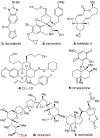
Structures of selected Golgi-disturbing agents.
Fig. 2
Fig. 2
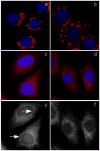
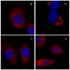
Phenotypic comparison of selected Golgi disturbing agents. NRK cells (a) were treated with the following Golgi disturbing agents: nocodazole (b), brefeldin A (c, e), ilimaquinone (d, f). Arrows indicate nuclear envelope staining. Golgi complex is shown in red, nuclei in blue.
Fig. 3
Fig. 3
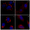
Norrisolide induces irreversible Golgi fragmentation. NRK cells plated on coverslips were treated with norrisolide for 0 (a) and 60 minutes (b). Cells were then washed and allowed to recover in fresh medium (c). Golgi complex is shown in red, nuclei in blue.
Fig. 4
Fig. 4
The norrisolide-induced Golgi fragmentation is independent on intact MT cytoskeleton and PKD activity. NRK cells pretreated for 60 min with nocodazole (a) were incubated with norrisolide for additional 60 min (b). NRK cells were treated for 60 min with IQ (c) or norrisolide (d) in presence of PKD inhibitor H89.
Fig. 5
Fig. 5
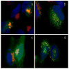
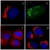
Protein transport in norrisolide-treated cells. Cells transfected with VSV-G (green) were incubated at 40 °C, then shifted to 20 °C (a, b) and 32 °C (c, d) in presence of DMSO (a, c) and norrisolide (b, d). VSV-G localizes in Golgi (a,b) and at the plasma membrane (c,d). Golgi complex is shown in red, VSV-G in green. Nuclei are in blue. Colocalization between Golgi and VSV-G is in yellow
Fig. 6
Fig. 6
Synthetic analogues of norrisolide
Fig. 7
Fig. 7
Effect of norrisolide analogue 8 on Golgi vesiculation. NRK cells were treated with 8 for 60 min (a) and allowed to recover for 60 min (b). Golgi complex is shown in red. Nuclei are in blue.
Fig. 8
Fig. 8
Norrisolide-based fluorescent probes
Fig. 9
Fig. 9
Fluorescent analogues of norrisolide induce Golgi fragmentation and localize on Golgi. NRK cells were treated with 15 (a) and 14 (b) for 60 min. Cells were treated with 14 for 10 min and imaged in live conditions (c). Fixed cells were labeled with a Golgi-specific antibody (d) then incubated with 14 (e); yellow color in (f) indicates colocalization of probe 14 and the antibody. Competition experiment: fixed NRK cells were incubated with 14 in presence of cell medium (g), DMSO (h) or 7 (i). Golgi complex is shown in red. Coumarin fluorescence is shown in green. Nuclei are in blue.
Fig. 10
Fig. 10
Norrisolide-based probes 17-20
Fig. 11
Fig. 11
Effect of probes 17 and 18 on Golgi. NRK cells were treated with the probe 17 for 60 min (a) and then allowed to recover for additional 60 min (b). Cells were treated with 18 for 60 min (c) and then allowed to recover (b). Golgi complex is shown in red. Nuclei are in blue.
Fig. 12
Fig. 12
A norrisolide-based trifunctional probe 21 that combines the perhydroindane core with an epoxide and a biotin motif.
Fig. 13
Fig. 13
Effect of probe 21 NRK on Golgi. Cells were treated with 21 for 60 min (a); then washed and incubated in fresh medium for 60 min (b). Asterisks indicate cells with fragmented Golgi. Golgi is shown in red; nuclei in blue.
Fig. 14
Fig. 14
A norrisolide-based trifunctional probe that combines the perhydroindane core with two epoxides and a DMACA fluorophore.
Fig. 15
Fig. 15
Effect of probe 22 on Golgi. Fixed NRK cells were labeled with Golgi-specific antibody (a); live cells were incubated with 22 for 10 min (b). Cells were treated with 22 for 60 min (c) and then allowed to recover for 60 min (d). Golgi complex is shown in red. DMACA fluorescence is shown in green. Nuclei are in blue.

Similar articles

Cited by

References

    1. For selected reviews on the secretory pathway see: Pelham HR. Cell Struct Funct. 1996;5:413–419.Rothman JE, Orci L. Nature. 1992;355:409–415.Pelham HR, Munro S. Cell. 1993;75:603–605.Corda D, Hidalgo Carcedo C, Bonazzi M, Luini A, Spanò S. Cell Mol Life Sci. 2002;59:1819–1832.Luini A, Mironov AA, De Matteis MA, Griffiths G. Med Secoli. 2007;19:29–54.Lowe M, Nakamura N, Warren G. Trends Cell Biol. 1998;8:40–44.

    1. For selected reports on this topic see: Fahy JV. Chest. 2002;122:320S–326S.Suchy SF, Olivos-Glander IM, Nussabaum RL. Hum Mol Genet. 1995;4:2245–2250.Cheng SH, Gregory RJ, Marshall J, Paul S, Souza DW, White GA, O'Riordan CR, Smith AE. Cell. 1990;63:827–834.

    1. For selected reports on this topic see: Spooner RA, Smith DC, Easton AJ, Roberts LM, Lord JM. Virol J. 2006;3:26.Sandvig K, van Deurs B. Annu Rev Cell Dev Biol. 2002;18:1–24.Ludwig JW, Richards ML. Curr Top Med Chem. 2006;6:165–178.

    1. For selected reports on this topic see: Roth J. Chem Rev. 2002;102:285–303.Arnold SM, Kaufman RJ. New Compreh Biochem. 2003;38:411–432.van Vliet C, Thomas EC, Merino-Trigo A, Teasdale RD, Gleeson PA. Progr Biophys & Mol Biol. 2003;83:1–45.Schulein R. Rev Physiol Biochem & Pharmacol. 2004;151:45–91.Sifers RN. Science. 2003;299:1330–1331.

    1. For selected reports on this topic see: Warren G, Malhotra V. Curr Opin Cell Biol. 1998;10:493–498.Bard F, Malhotra V. Annu Rev Cell Dev Biol. 2006;22:439–455.

Publication types

LinkOut - more resources