. 2010 Apr;14(4):147-53.
doi: 10.1016/j.tics.2010.01.007. Epub 2010 Feb 26.

Visual stability based on remapping of attention pointers

Affiliations

Visual stability based on remapping of attention pointers

Patrick Cavanagh et al. Trends Cogn Sci. 2010 Apr.

Abstract

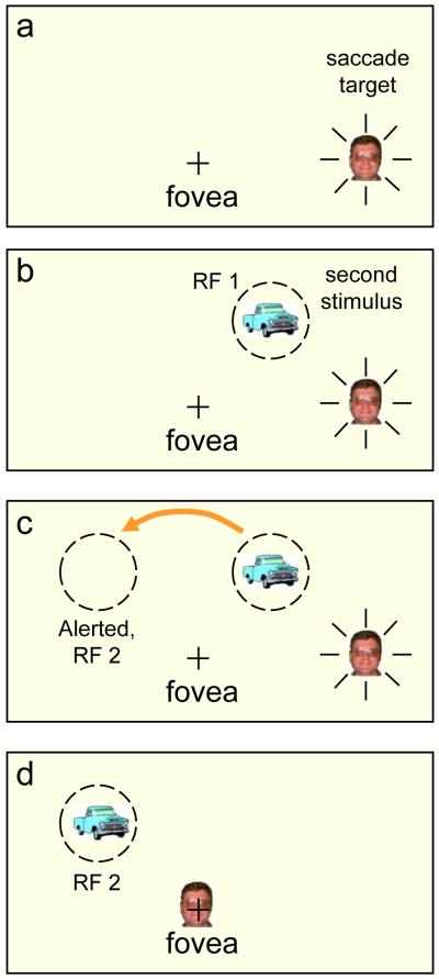
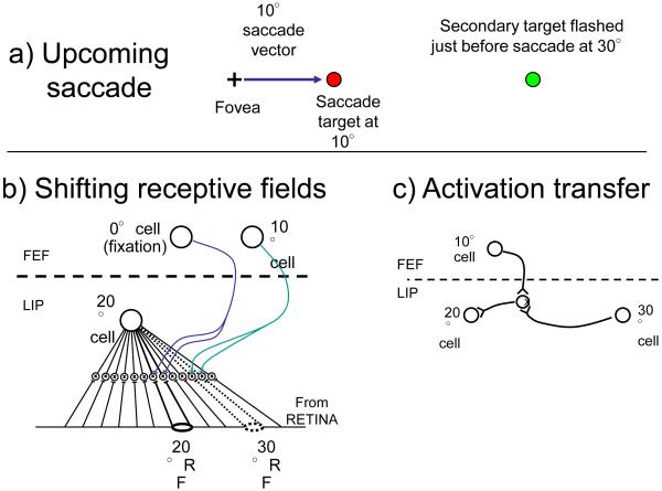
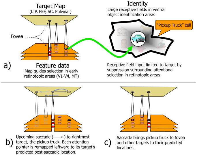
When we move our eyes, we easily keep track of where relevant things are in the world. Recent proposals link this stability to the shifting of receptive fields of neurons in eye movement and attention control areas. Reports of 'spatiotopic' visual aftereffects have also been claimed to support this shifting connectivity even at an early level, but these results have been challenged. Here, the process of updating visual location is described as predictive shifts of location 'pointers' to attended targets, analogous to predictive activation seen cross-modally. We argue that these location pointers, the core operators of spatial attention, are linked to identity information and that such a link is necessary to establish a workable visual architecture and to explain frequently reported positive spatiotopic biases.


Figures

Figure 1
Figure 1. Remapping
a) the “+” indicates the fovea and a face is the target of an impending saccade. b) A second stimulus is flashed on just before the saccade, activating a cell (in LIP) with a receptive field, RF1, at the location of the new stimulus. c) The activation of the cell with classic receptive field on the right is transferred to a new cell whose receptive field, RF 2, is on the left; critically, its receptive field lies where the second stimulus will land after the saccade. This cell’s predictive activation gives a head start for the processing of this stimulus when it lands. d) The saccade has landed on the face and the truck lands in the pre-activated receptive field. In the physiological experiments [e.g., 13] the second stimulus is extinguished before the saccade lands so that any response from RF2 can be attributed solely to the pre-saccadic activity at the remote location and to the intended saccade and its corollary discharge or efference copy.
Figure 2
Figure 2. Simplified examples of two remapping mechanisms
a) An upcoming saccade to a target at 10° with a briefly presented second target at 30°. This will activate some cells with classic receptive fields at 20° even though the secondary target does not land there until after the saccade. b) Shifting field. Each cell in a salience map (shown as LIP here) is connected to all locations on the retina through interneurons. Input from an oculomotor center (FEF here) turns on subsets of the input that correspond to the default location when fixating (saccade of 0°) or the appropriately offset location when a saccade is imminent (10° here). The shifted location to be activated depends on the cell and the saccade. The cell shown with its classical receptive field at 20° will now respond to the input at 30°. c) Activation transfer. Horizontal connections link all cells on a salience map (LIP here). An upcoming saccade will open all connections of the appropriate offset, transferring any activation from cells with pre-saccadic targets to locations where those targets will fall after the saccade. These connections must be learned [30, 31]. Here the secondary target activates the cell with receptive field at 30° and this activity is transferred to the 20° cell through the active interneuron.
Figure 3
Figure 3. Remapping and attention
A network of areas form a target map that subserves spatial attention as well as eye movements [26-28]. a) Peaks of activity (in red) index the locations of targets and specify the retinotopic coordinates at which the target’s feature data are to be found in earlier visual cortices which are shown, highly simplified, as a stack of aligned areas divided into right and left hemifields with the fovea in the center. In object recognition areas, cells have very large receptive fields (heavy black outline) and depend on attention to bias input [67] in favor of the target and suppress surrounding distractors so that only a single item falls in the receptive field at any one time. b) Just around the time of a saccade, the activity peaks for the 3 attended targets shift to the locations the targets will have at the end of the saccade, (c) and, following the saccade, what had been the saccade target, at the right, now lands in the fovea.

Comment in

References

    1. Breitmeyer BG, et al. The existence and role of retinotopic and spatiotopic forms of visual persistence. Acta Psychol (Amst) 1982;52:175–196. - PubMed
    1. Duhamel JR, et al. Spatial invariance of visual receptive fields in parietal cortex neurons. Nature. 1997;389:845–848. - PubMed
    1. Galletti C, et al. Parietal neurons encoding spatial locations in craniotopic coordinates. Exp Brain Res. 1993;96:221–229. - PubMed
    1. Melcher D. Spatiotopic transfer of visual-form adaptation across saccadic eye movements. Curr Biol. 2005;15:1745–1748. - PubMed
    1. Melcher D, Morrone MC. Spatiotopic temporal integration of visual motion across saccadic eye movements. Nat Neurosci. 2003;6:877–881. - PubMed

Publication types