Review
. 2010 May;135(2):169-82.
doi: 10.1016/j.clim.2010.01.011. Epub 2010 Mar 1.

Immunodeficiency due to mutations in ORAI1 and STIM1

Affiliations
Review

Immunodeficiency due to mutations in ORAI1 and STIM1

Stefan Feske et al. Clin Immunol. 2010 May.

Abstract

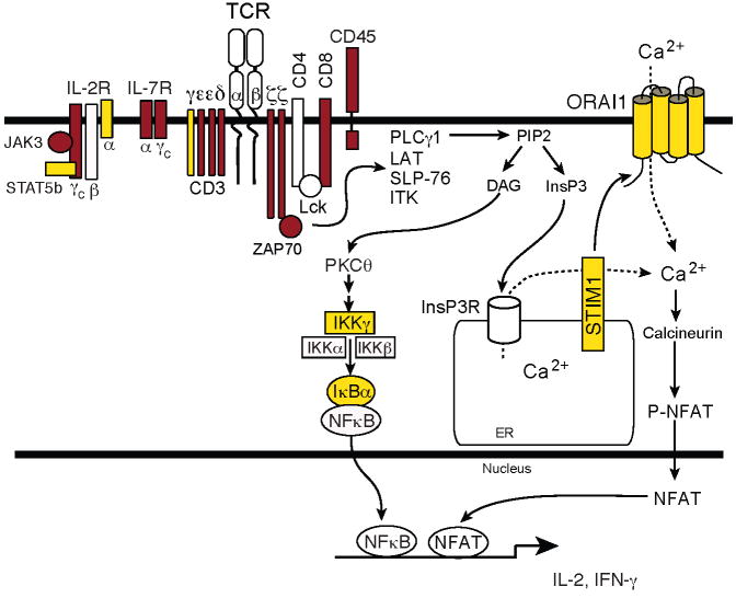
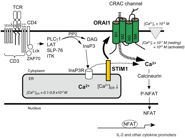
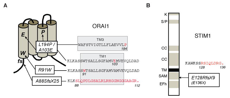
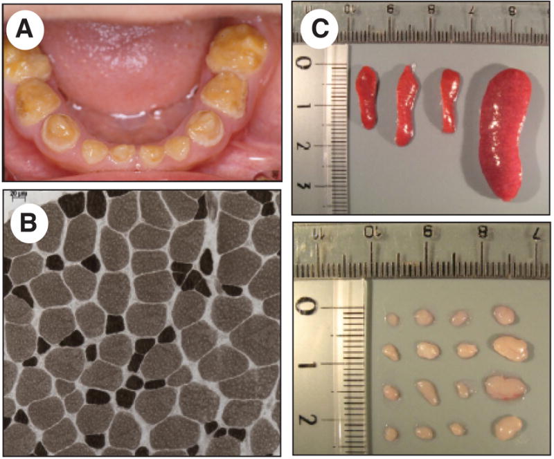
Lymphocyte activation requires Ca(2+) influx through specialized Ca(2+) channels in the plasma membrane. In T cells the predominant Ca(2+) channel is the Ca(2+) release activated Ca(2+) (CRAC) channel encoded by the gene ORAI1. ORAI1 is activated by stromal interaction molecule (STIM) 1 that is localized in the ER where it senses the concentration of stored Ca(2+). Following antigen binding to immunoreceptors such as the TCR, ER Ca(2+) stores are depleted, STIM1 is activated and ORAI1-CRAC channels open resulting in what is referred to as store-operated Ca(2+) entry (SOCE). Mutations in ORAI1 and STIM1 genes in human patients that lead to expression of non-functional ORAI1 or complete lack of ORAI1 or STIM1 protein are associated with a unique clinical phenotype that is characterized by immunodeficiency, muscular hypotonia and anhydrotic ectodermal dysplasia, as well as, in the case of STIM1 deficiency, autoimmunity and lymphoproliferative disease. The immunodeficiency in these patients is due to a severe defect in T cell activation but not in lymphocyte development. This review describes the immunological and non-immunological phenotypes of patients with defects in SOCE and CRAC channel function and discusses them in the context of similar immunodeficiency diseases and animal models of ORAI1 and STIM1 function.


Figures

Figure 1
Figure 1. Molecules mutated in SCID and T cell activation defects
Patients with mutations in ORAI1, STIM1, CD3γ and IκBα (yellow symbols) have impaired T cell activation and but normal numbers of lymphocytes. By contrast, SCID patients with mutations in other signaling molecules (red symbols) show severe defects in the development of T cells or T cell subsets [2]. Abbreviations: DAG, diacylglycerol; InsP3, inositol-1,4,5-trisphosphate; LAT, linker for activation of T cells; NFAT, nuclear factor of activated T cells; PLC, phospholipase; PtdIns(4,5)P2, phosphatidylinositol-4,5-bisphosphate; SLP76, SH2-domain-containing leukocyte protein of 76 kDa; STIM1, stromal interaction molecule 1; ZAP-70, ζ-chain-associated protein kinase of 70 kDa.
Figure 2
Figure 2. Store-operated Ca2+ entry (SOCE) in T cells
T-cell receptor (TCR) engagement results in the activation of tyrosine kinases Lck and ZAP-70, assembly of the adaptor protein complex containing SLP76 and LAT and activation of PLCγ1. The latter hydrolyses PtdIns(4,5)P2 to InsP3 and DAG. InsP3 binds to and opens InsP3 receptors (InsP3Rs) in the ER, resulting in the release of Ca2+ from ER stores, reduction of [Ca2+]ER and transient increase in [Ca2+]i. The descrease in [Ca2+]ER is sensed by STIM1 resulting in binding of STIM1 to ORAI1 and opening of the CRAC channel. Ca2+ influx results in increased intracellular Ca2+ concentration [Ca2+]i from ~ 100 nM to ~1 μM. Sustained elevation of [Ca2+]i is required for activation of the phosphatase calcineurin, nuclear translocation of the transcription factor NFAT and cytokine gene expression. For abbreviations see Figure 1.
Figure 3
Figure 3. Phenotypic detection of putative heterozygous carriers
Heterozygous carriers of an autosomal recessive mutation that impairs Ca2+ influx in immunodeficient patients were predicted based on in vitro analysis of SOCE in T cells from relatives of immunodeficient patients (see pedigree in Figure 4A). A, Ca2+ influx was measured in T cells stimulated with thapsigargin (TG) followed by readdition of lower than physiological extracellular Ca2+ (0.5 mM, black bar) to reveal a defect in Ca2+ influx. B, T cells from immunodeficient patients (purple trace in A and bar in B) lack SOCE, whereas T cells from relatives have normal (blue) or markedly reduced (green) Ca2+ influx, indicating that the latter, although healthy, may be heterozygous carriers of a recessive, disease-causing mutation. Not shown, The haplotypes of immunodeficient patients, predicted heterozygotes and predicted wild-type individuals from the same family were determined by genome-wide single nucleotide polymorphism (SNP) analysis and used to calculate multipoint parametric LOD scores in two independent analyses assuming an autosomal recessive and autosomal dominant mode of inheritance, respectively [17]. A 6.5 Mb interval on chromosome 12q24 containing the novel gene ORAI1 was identified to be linked to immunodeficiency disease with a LOD (log10 of the odds) score of 5.7. Identification of heterozygous carriers may be a useful tool for identification of gene defects in other rare diseases because it increases the number of haplotypes available for linkage analysis. DNA sequence analysis of ORAI1 revealed that all predicted carriers were indeed heterozygous for the ORAI1-R91W mutation. Figure modified from the version originally published in [17].
Figure 4
Figure 4. Pedigrees of patients lacking SOCE
ORAI1-R91W patients were first reported in [17]. ORAI1-A88SfsX25 and ORAI1-A103E/L194P patients were first reported in [33](pedigrees reproduced with permission). STIM1-E128RfsX9 (E136X) patients were first reported in [19](pedigree reproduced with permission). Black symbols depict patients, strike-through symbols deceased individuals, dotted symbols heterozygous carriers. All heterozygous carriers are healthy.
Figure 5
Figure 5. Mutations in ORAI1 and STIM1 in immunodeficient patients lacking Ca2+ influx
A, ORAI1 is a plasma membrane Ca2+ channel with four transmembrane domains and intracellular N- and C-termini (for details see text). Mutations: (i) An Arg→Trp (R91W) single amino acid substitution in ORAI1 at the beginning of the first transmembrane (TM1) domain results in expression of a nonfunctional protein [17]. (ii) A frameshift (fs) nonsense mutation in the first exon of ORAI1 (A88SfsX25) results in premature termination, nonsense mediated decay of ORAI1 mRNA and abolished ORAI1 protein expression [33]. (iii) Two independent single amino acid substitutions, A103E and L194P, in TM1 and TM3, respectively interfere with ORAI1 protein expression [33]. B, STIM1 is a single-pass transmembrane (TM) protein localized in the ER featuring an EF hand domain (EFh), sterile alpha motif (SAM), coiled-coil (CC), serin/proline (S/P) and lysine (K) rich domains (for details see text). Mutation: A frameshift nonsense mutation in exon 3 of STIM1 (E128RfsX9) results in premature termination, nonsense mediated mRNA decay of STIM1 and abolished STIM1 protein expression [19].
Figure 6
Figure 6. Non-immunological phenotypes in patients and mice lacking SOCE
A, Amelogenesis imperfecta type III in a patient with ORAI1-R91W mutation. The dental enamel is hypocalcified resulting in use-dependent loss of the enamel layer. Shown are deciduous teeth at 6 years of age. B, Muscular dysplasia in a patient with ORAI1-R91W mutation. Atrophic type II muscle fibers in ATPase staining of a muscle biopsy at age 5 years (reproduced with permission from [33]. C, Splenomegaly and lymphadneopathy in Stim1f/f Stim2f/f Cd4-Cre mice [58]. Mice also show leukocytic infiltration of lung and liver due to reduced numbers and impaired function of Foxp3+ regulatory T cells. A similar lymphoproliferative phenotype was observed in patients with STIM1 mutation [19].

References

    1. Fischer A. Human Primary Immunodeficiency Diseases. Immunity. 2007;27:835–845. - PubMed
    1. Notarangelo LD, Fischer A, Geha RS, Casanova JL, Chapel H, Conley ME, Cunningham-Rundles C, Etzioni A, Hammartrom L, Nonoyama S, Ochs HD, Puck J, Roifman C, Seger R, Wedgwood J. Primary immunodeficiencies: 2009 update. J Allergy Clin Immunol. 2009;124:1161–78. - PMC - PubMed
    1. Buckley RH. Molecular defects in human severe combined immunodeficiency and approaches to immune reconstitution. Annu Rev Immunol. 2004;22:625–655. - PubMed
    1. Castigli E, Pahwa R, Good RA, Geha RS, Chatila TA. Molecular basis of a multiple lymphokine deficiency in a patient with severe combined immunodeficiency. Proc Natl Acad Sci U S A. 1993;90:4728–32. - PMC - PubMed
    1. Chatila T, Castigli E, Pahwa R, Pahwa S, Chirmule N, Oyaizu N, Good RA, Geha RS. Primary combined immunodeficiency resulting from defective transcription of multiple T-cell lymphokine genes. Proc Natl Acad Sci U S A. 1990;87:10033–7. - PMC - PubMed

Publication types