

The role of amygdala nuclei in the expression of auditory signaled two-way active avoidance in rats
- PMID: 20189958
- PMCID: PMC2832923
- DOI: 10.1101/lm.1676610
The role of amygdala nuclei in the expression of auditory signaled two-way active avoidance in rats
Abstract
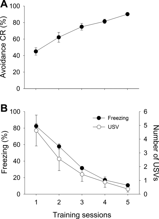
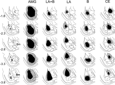
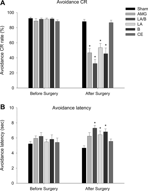
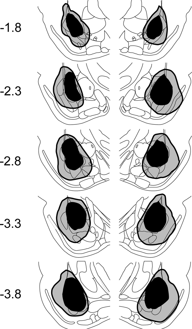
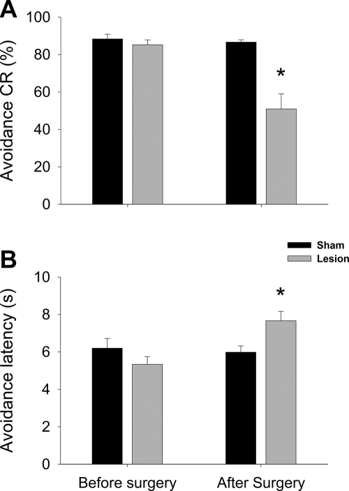
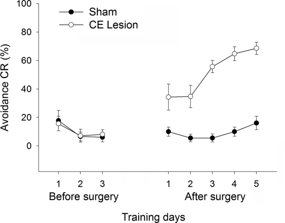
Using a two-way signaled active avoidance (2-AA) learning procedure, where rats were trained in a shuttle box to avoid a footshock signaled by an auditory stimulus, we tested the contributions of the lateral (LA), basal (B), and central (CE) nuclei of the amygdala to the expression of instrumental active avoidance conditioned responses (CRs). Discrete or combined lesions of the LA and B, performed after the rats had reached an asymptotic level of avoidance performance, produced deficits in the CR, whereas CE lesions had minimal effect. Fiber-sparing excitotoxic lesions of the LA/B produced by infusions of N-methyl-d-aspartate (NMDA) also impaired avoidance performance, confirming that neurons in the LA/B are involved in mediating avoidance CRs. In a final series of experiments, bilateral electrolytic lesions of the CE were performed on a subgroup of animals that failed to acquire the avoidance CR after 3 d of training. CE lesions led to an immediate rescue of avoidance learning, suggesting that activity in CE was inhibiting the instrumental CR. Taken together, these results indicate that the LA and B are essential for the performance of a 2-AA response. The CE is not required, and may in fact constrain the instrumental avoidance response by mediating the generation of competing Pavlovian responses, such as freezing.
Figures
Similar articles
-
Sidman instrumental avoidance initially depends on lateral and basal amygdala and is constrained by central amygdala-mediated Pavlovian processes.Biol Psychiatry. 2010 Jun 15;67(12):1120-7. doi: 10.1016/j.biopsych.2009.12.002. Epub 2010 Jan 27. Biol Psychiatry. 2010. PMID: 20110085 Free PMC article.
-
Active avoidance learning requires prefrontal suppression of amygdala-mediated defensive reactions.J Neurosci. 2013 Feb 27;33(9):3815-23. doi: 10.1523/JNEUROSCI.2596-12.2013. J Neurosci. 2013. PMID: 23447593 Free PMC article.
-
Neurotoxic lesions of the lateral nucleus of the amygdala decrease conditioned fear but not unconditioned fear of a predator odor: comparison with electrolytic lesions.J Neurosci. 2001 May 15;21(10):3619-27. doi: 10.1523/JNEUROSCI.21-10-03619.2001. J Neurosci. 2001. PMID: 11331391 Free PMC article.
-
The amygdala modulates memory consolidation of fear-motivated inhibitory avoidance learning but not classical fear conditioning.J Neurosci. 2000 Sep 15;20(18):7059-66. doi: 10.1523/JNEUROSCI.20-18-07059.2000. J Neurosci. 2000. PMID: 10995852 Free PMC article.
-
Conditioned taste aversion and amygdala lesions in the rat: a critical review.Neurosci Biobehav Rev. 2005;29(7):1067-88. doi: 10.1016/j.neubiorev.2005.03.025. Neurosci Biobehav Rev. 2005. PMID: 15893375 Review.
Cited by
-
Active vs. reactive threat responding is associated with differential c-Fos expression in specific regions of amygdala and prefrontal cortex.Learn Mem. 2013 Jul 18;20(8):446-52. doi: 10.1101/lm.031047.113. Learn Mem. 2013. PMID: 23869027 Free PMC article.
-
Circuits That Mediate Expression of Signaled Active Avoidance Converge in the Pedunculopontine Tegmentum.J Neurosci. 2019 Jun 5;39(23):4576-4594. doi: 10.1523/JNEUROSCI.0049-19.2019. Epub 2019 Apr 1. J Neurosci. 2019. PMID: 30936242 Free PMC article.
-
Rate and Temporal Coding Mechanisms in the Anterior Cingulate Cortex for Pain Anticipation.Sci Rep. 2018 May 29;8(1):8298. doi: 10.1038/s41598-018-26518-x. Sci Rep. 2018. PMID: 29844413 Free PMC article.
-
D-Serine as the gatekeeper of NMDA receptor activity: implications for the pharmacologic management of anxiety disorders.Transl Psychiatry. 2020 Jun 9;10(1):184. doi: 10.1038/s41398-020-00870-x. Transl Psychiatry. 2020. PMID: 32518273 Free PMC article. Review.
-
Lesions of lateral or central amygdala abolish aversive Pavlovian-to-instrumental transfer in rats.Front Behav Neurosci. 2014 May 7;8:161. doi: 10.3389/fnbeh.2014.00161. eCollection 2014. Front Behav Neurosci. 2014. PMID: 24847229 Free PMC article.
References
-
- Amorapanth P, LeDoux JE, Nader K. Different lateral amygdala outputs mediate reactions and actions elicited by a fear-arousing stimulus. Nat Neurosci. 2000;3:74–79. - PubMed
-
- Antoniadis EA, McDonald RJ. Discriminative fear conditioning to context expressed by multiple measures of fear in the rat. Behav Brain Res. 1999;101:1–13. - PubMed
-
- Blanchard RJ, Blanchard DC. Defensive reactions in the albino rat. Learn Motivat. 1971;2:351–362.
-
- Brennan FX, Beck KD, Servatius RJ. Lever press escape/avoidance conditioning in rats: Safety signal length and avoidance performance. Integr Physiol Behav Sci. 2003;38:36–44. - PubMed
Publication types
MeSH terms
Grants and funding
LinkOut - more resources
Full Text Sources
Miscellaneous