. 2010 May;192(9):2373-84.
doi: 10.1128/JB.01546-09. Epub 2010 Feb 26.

Characterization of Acp, a peptidoglycan hydrolase of Clostridium perfringens with N-acetylglucosaminidase activity that is implicated in cell separation and stress-induced autolysis

Affiliations

Characterization of Acp, a peptidoglycan hydrolase of Clostridium perfringens with N-acetylglucosaminidase activity that is implicated in cell separation and stress-induced autolysis

Emilie Camiade et al. J Bacteriol. 2010 May.

Abstract

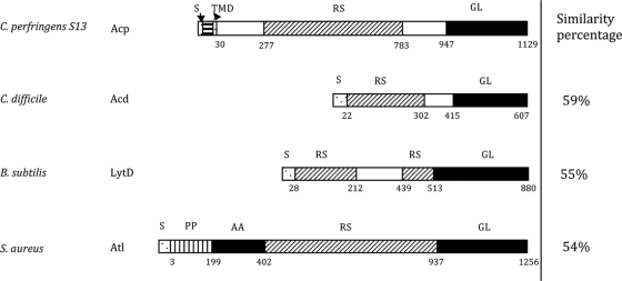
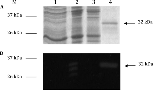
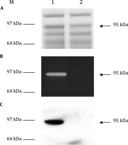
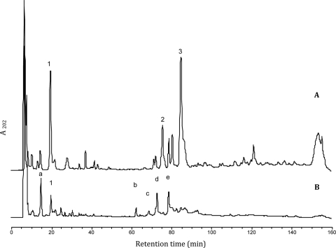
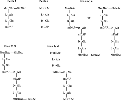
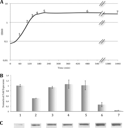
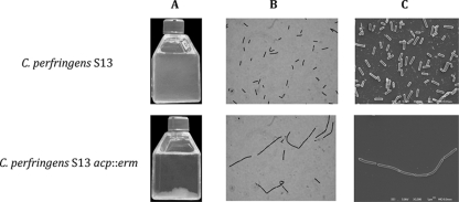
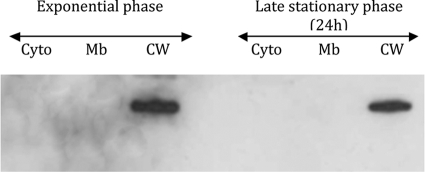
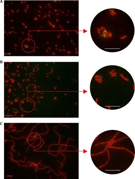
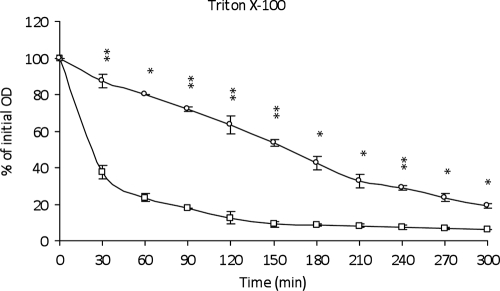
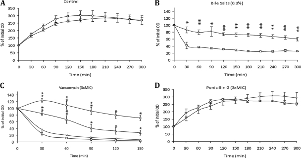
This work reports the characterization of the first known peptidoglycan hydrolase (Acp) produced mainly during vegetative growth of Clostridium perfringens. Acp has a modular structure with three domains: a signal peptide domain, an N-terminal domain with repeated sequences, and a C-terminal catalytic domain. The purified recombinant catalytic domain of Acp displayed lytic activity on the cell walls of several Gram-positive bacterial species. Its hydrolytic specificity was established by analyzing the Bacillus subtilis peptidoglycan digestion products by coupling reverse phase-high-pressure liquid chromatography (RP-HPLC) and matrix-assisted laser desorption ionization-time of flight mass spectrometry (MALDI-TOF MS) analysis, which displayed an N-acetylglucosaminidase activity. The study of acp expression showed a constant expression during growth, which suggested an important role of Acp in growth of C. perfringens. Furthermore, cell fractionation and indirect immunofluorescence staining using anti-Acp antibodies revealed that Acp is located at the septal peptidoglycan of vegetative cells during exponential growth phase, indicating a role in cell separation or division of C. perfringens. A knockout acp mutant strain was obtained by using the insertion of mobile group II intron strategy (ClosTron). The microscopic examination indicated a lack of vegetative cell separation in the acp mutant strain, as well as the wild-type strain incubated with anti-Acp antibodies, demonstrating the critical role of Acp in cell separation. The comparative responses of wild-type and acp mutant strains to stresses induced by Triton X-100, bile salts, and vancomycin revealed an implication of Acp in autolysis induced by these stresses. Overall, Acp appears as a major cell wall N-acetylglucosaminidase implicated in both vegetative growth and stress-induced autolysis.


Figures

FIG. 1.
FIG. 1.
Modular organization of C. perfringens Acp compared to C. difficile Acd, B. subtilis LytD, and S. aureus Atl. Percent similarity between the catalytic domain of Acp (black rectangles) and the three other autolysins are indicated to the right. S, signal sequence; RS, repeated sequence; GL, N-acetylglucosaminidase; AA, l-alanyl-amidase; TMD, transmembrane domain.
FIG. 2.
FIG. 2.
Purification of His-tagged Acp catalytic domain. Analysis of protein extracts on SDS-PAGE (A) and renaturing SDS-PAGE (B) containing 0.2% Micrococcus lysodeikticus cell wall (zymogram). Lane 1, crude cell extract of E. coli BL21 carrying pET28; lane 2, crude cell extract of E. coli BL21 carrying pCD470 induced by 1 mM IPTG; lane 3, crude cell extract of E. coli BL21 carrying noninduced pCD470; and lane 4, purified His-tagged Acp catalytic domain under native conditions. M, benchmark protein ladders (Invitrogen).
FIG. 3.
FIG. 3.
Detection of Acp in the total crude extract of C. perfringens strain 13 (lane 1) and C. perfringens strain 13 acp::erm (lane 2). (A) Coomassie blue-stained SDS-PAGE. (B) Methylene blue-stained zymogram containing M. lysodeikticus lyophilized cells. (C) Western blot. M, molecular mass marker (SeeBlue Plus 2 pre-stained standard; Invitrogen).
FIG. 4.
FIG. 4.
RP-HPLC analysis of the soluble muropeptides released from B. subtilis vegetative peptidoglycan after incubation with Acp (A) or with Acp and mutanolysin (B). The numbers and letters indicate the peaks analyzed by MALDI-TOF MS.
FIG. 5.
FIG. 5.
Structure of the muropeptides from B. subtilis peptidoglycan obtained after Acp digestion (peaks 1, 2, and 3) or Acp plus mutanolysin digestion (peaks a to e). Peak numbers or letters refer to the peaks on the chromatograms presented in Fig. 4. According to their mass (Table 1), peaks 2, b, and c bear one amidation whereas peaks 3, d, and e bear two amidations, located most probably on mDAP (2).
FIG. 6.
FIG. 6.
Analysis of acp during growth of C. perfringens strain 13 (BHI medium at 37°C under anaerobic conditions). (A) Growth of C. perfringens strain 13 followed by OD600; numbers 1 to 7 represent the different times of total RNA and protein preparation used for qRT-PCR and Western blot analysis. (B) qRT-PCR analysis results showing relative expression of acp normalized by the housekeeping gene 16S rRNA during the different growth phases of C. perfringens strain 13 and compared to the early exponential phase. Error bars indicate standard deviation. (C) Western blot of Acp with polyclonal anti-Acp antibodies.
FIG. 7.
FIG. 7.
Macroscopic and microscopic observations of C. perfringens strain 13 and C. perfringens strain 13 acp::erm. (A) Overnight BHI broth culture. (B) Gram staining microscopy view. (C) Scanning electron microscopy.
FIG. 8.
FIG. 8.
Cell fractionation localization of Acp during exponential and late stationary growth phases of C. perfringens as follows: cytoplasm (Cyto), membrane (Mb), and cell wall (CW). Protein extracts were analyzed by Western blotting with specific anti-Acp antibodies.
FIG. 9.
FIG. 9.
Immunolocalization of Acp on C. perfringens strain 13 during exponential growth phase by indirect immunofluorescence. Bar, 10 μM. Red staining fluorescence is due to the monomeric cyanin nucleic acid stain To-Pro-3, and green fluorescence is due to the secondary anti-mouse IgG coupled with Alexa Fluor 488 fluorophore. (A) C. perfringens strain 13 stained with depleted anti-Acp immune serum. (B) C. perfringens strain 13 stained with depleted anti-Acp preimmune serum as a control. (C) C. perfringens strain 13 acp::erm stained with depleted anti-Acp immune serum.
FIG. 10.
FIG. 10.
Autolytic activities of C. perfringens strain 13 during Triton X-100-induced autolysis. The autolysis is expressed in percent initial absorbance at an optical density of 600 nm. Shown are results for C. perfringens strain 13 (□) and C. perfringens strain 13 acp::erm (○). Error bars indicate standard deviations, and asterisks indicate statistically significant differences (*, P < 0.05; **, P < 0.01).
FIG. 11.
FIG. 11.
Impact of acp inactivation on stress-induced autolysis of C. perfringens strain 13. Growth of mid-exponential phase cultures was determined at 37°C in the absence (A) or presence (B) of 0.3% of bile salts, 3× MIC of vancomycin (C), or 3× MIC of penicillin G (D) by measuring the optical densities (expressed as percent initial absorbance at an optical density at 600 nm). Shown are results for C. perfringens strain 13 (□), C. perfringens strain 13 acp::erm (○), C. perfringens strain 13 plus preimmune serum (×), and C. perfringens strain 13 plus polyclonal anti-Acp antibodies (▵). Error bars indicate standard deviations, and asterisks indicate statistically significant differences compared to C. perfringens strain 13 (*, P < 0.05; **, P < 0.01).
FIG. 12.
FIG. 12.
Effect of anti-Acp polyclonal antibodies on chain length. C. perfringens strain 13 was grown in BHI broth including polyclonal anti-Acp antibody or preimmune serum (1/50).

Similar articles

Cited by

References

    1. Allignet, J., P. England, I. Old, and N. El Solh. 2002. Several regions of the repeat domain of the Staphylococcus caprae autolysin, AtlC, are involved in fibronectin binding. FEMS Microbiol. Lett. 213:193-197. - PubMed
    1. Atrih, A., G. Bacher, G. Allmaier, M. P. Williamson, and S. J. Foster. 1999. Analysis of peptidoglycan structure from vegetative cells of Bacillus subtilis 168 and role of PBP 5 in peptidoglycan maturation. J. Bacteriol. 181:3956-3966. - PMC - PubMed
    1. Bateman, A., and M. Bycroft. 2000. The structure of a LysM domain from E. coli membrane-bound lytic murein transglycosylase D (MltD). J. Mol. Biol. 299:1113-1119. - PubMed
    1. Berry, A. M., R. A. Lock, D. Hansman, and J. C. Paton. 1989. Contribution of autolysin to virulence of Streptococcus pneumoniae. Infect. Immun. 57:2324-2330. - PMC - PubMed
    1. Bourgeois, I., E. Camiade, R. Biswas, P. Courtin, L. Gibert, F. Gotz, M. P. Chapot-Chartier, J. L. Pons, and M. Pestel-Caron. 2009. Characterization of AtlL, a bifunctional autolysin of Staphylococcus lugdunensis with N-acetylglucosaminidase and N-acetylmuramoyl-l-alanine amidase activities. FEMS Microbiol. Lett. 290:105-113. - PubMed

Publication types

MeSH terms

Associated data

LinkOut - more resources