. 2010 May;192(9):2346-58.
doi: 10.1128/JB.01690-09. Epub 2010 Feb 26.

Kinetic characterization of the WalRKSpn (VicRK) two-component system of Streptococcus pneumoniae: dependence of WalKSpn (VicK) phosphatase activity on its PAS domain

Affiliations

Kinetic characterization of the WalRKSpn (VicRK) two-component system of Streptococcus pneumoniae: dependence of WalKSpn (VicK) phosphatase activity on its PAS domain

Alina D Gutu et al. J Bacteriol. 2010 May.

Abstract

The WalRK two-component system plays important roles in maintaining cell wall homeostasis and responding to antibiotic stress in low-GC Gram-positive bacteria. In the major human pathogen, Streptococcus pneumoniae, phosphorylated WalR(Spn) (VicR) response regulator positively controls the transcription of genes encoding the essential PcsB division protein and surface virulence factors. WalR(Spn) is phosphorylated by the WalK(Spn) (VicK) histidine kinase. Little is known about the signals sensed by WalK histidine kinases. To gain information about WalK(Spn) signal transduction, we performed a kinetic characterization of the WalRK(Spn) autophosphorylation, phosphoryltransferase, and phosphatase reactions. We were unable to purify soluble full-length WalK(Spn). Consequently, these analyses were performed using two truncated versions of WalK(Spn) lacking its single transmembrane domain. The longer version (Delta35 amino acids) contained most of the HAMP domain and the PAS, DHp, and CA domains, whereas the shorter version (Delta195 amino acids) contained only the DHp and CA domains. The autophosphorylation kinetic parameters of Delta35 and Delta195 WalK(Spn) were similar [K(m)(ATP) approximately 37 microM; k(cat) approximately 0.10 min(-1)] and typical of those of other histidine kinases. The catalytic efficiency of the two versions of WalK(Spn) approximately P were also similar in the phosphoryltransfer reaction to full-length WalR(Spn). In contrast, absence of the HAMP-PAS domains significantly diminished the phosphatase activity of WalK(Spn) for WalR(Spn) approximately P. Deletion and point mutations confirmed that optimal WalK(Spn) phosphatase activity depended on the PAS domain as well as residues in the DHp domain. In addition, these WalK(Spn) DHp domain and DeltaPAS mutations led to attenuation of virulence in a murine pneumonia model.


Figures

FIG. 1.
FIG. 1.
Domain organization of the protein constructs used in the present study. Full-length WalKSpn (VicK) (line 1) contains 449 amino acids organized into five architectural and functional domains based on the SMART database (smart.embl-heidelberg.de): TM (anchoring transmembrane domain; amino acids 13 to 32), HAMP linker domain (amino acids 16 to 84), PAS domain consisting of PAS and PAC motifs (amino acids 94 to 202), DHp (dimerization histidine phosphoryltransfer [HisKA]; amino acids 208 to 274), and CA (kinase catalytic domain [HATPase]; amino acids 323 to 435). Histidine residue 218 (H218) is phosphorylated in the autokinase reaction. Numbering of full-length WalKSpn was extended to the soluble, truncated WalKSpn derivatives purified and characterized in the present study (lines 2 to 10; Materials and Methods; see Fig. S1 in the supplemental material). The affinity tags on the constructs are indicated. Full-length WalRSpn (VicR) contains 234 amino acids organized into two domains: a receiver domain (amino acids 2 to 112) and an effector domain (amino acids 154 to 230). Aspartate residue 52 in the receiver domain is phosphorylated in the transferase reaction with WalKSpn∼P constructs, and the effector domain contains the helix-turn-helix DNA binding motif. See the text for further details.
FIG. 2.
FIG. 2.
Progress curves of autophosphorylation reactions. Representative curves used to determine the kinetic parameters in Table 1 are shown. (A) Time course used to calculate the initial rates of WalKSpnΔN35 (C)-His autophosphorylation (Fig. 1, line 2) at different ATP concentrations. (B) Velocity versus [ATP] curve based on A used to calculate Km(ATP) and kcat for WalKSpnΔN35 (C)-His autophosphorylation. See Materials and Methods for details. These reactions contained 1.1 μM WalKSpnΔN35 (C)-His in a volume of 100 μl at 25°C.
FIG. 3.
FIG. 3.
WalKSpn∼P disappearance due to phosphoryltransfer to added WalRSpn. A representative reaction progression curve used to determine half-lives in Table 2 is shown. Open symbols, WalKSpnΔN35 (C)-His∼P decay in the absence of WalRSpn in reaction mixtures containing Mg2+ or Ca2+; closed symbols, decay of WalKSpnΔN35 (C)-His∼P in parallel reaction mixtures containing Mg2+ or Ca2+ and WalRSpn added at t = 0. See Materials and Methods for details. These reactions contained 1.5 μM WalKSpnΔN35 (C)-His∼P and 0.25 μM WalRSpn in a reaction volume of 100 μl at 25°C.
FIG. 4.
FIG. 4.
Autoradiographs showing autophosphorylation of WalKSpn Δ35 and Δ195 constructs, phosphoryltransfer to WalRSpn, and WalKSpn phosphatase of WalRSpn∼P. Representative time courses are shown and quantitated in Fig. S3 and S4 in the supplemental material. Combined reactions of WalKSpn autophosphorylation and WalRSpn phosphoryltransfer were performed at 25°C in reaction mixtures containing Mg2+ or Ca2+ as described in Materials and Methods. WalKSpn autophosphorylation reactions proceeded for 3 min before WalRSpn was added without removal of ATP (t = 0). Reactions contained the following concentrations of proteins: 2.2 μM WalKSpnΔN35 (C)-His (A), 2.9 μM WalKSpnΔN195 (C)-His (B), 3.4 μM WalKSpnΔN35 (N)-Sumo (C), and 2.6 μM WalKSpnΔN195 (N)-Sumo (D). Each reaction contained 9.6 μM WalRSpn.
FIG. 5.
FIG. 5.
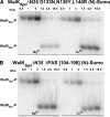
Autoradiographs showing autophosphorylation of WalKSpn PAS domain mutant constructs, phosphoryltransfer to WalRSpn, and WalKSpn phosphatase of WalRSpn∼P. Representative time courses are shown and quantitated in Fig. S5. Combined reactions of WalKSpn autophosphorylation and WalRSpn phosphoryltransfer were performed at 25°C in reaction mixtures containing Mg2+ or Ca2+ as described in Materials and Methods. WalKSpn autophosphorylation reactions proceeded for 3 min before WalRSpn was added without removal of ATP (t = 0). Reactions contained the following concentrations of proteins: 3.4 μM WalKSpnΔN35 D133N,N136Y,L140R (N)-Sumo (A) and 2.5 μM WalKSpnΔN35 ΔPAS[104-198] (N)-Sumo (B). Each reaction contained 9.6 μM WalRSpn.
FIG. 6.
FIG. 6.
WalKSpn phosphatase activity of WalRSpn∼P. Representative reaction progression curves used to determine the rate constants and half-lives in Tables 4 and 5 are shown. Reactions containing 13.2 μM ADP were carried out at 25°C as described in Materials and Methods. (A) Reversed-phase HPLC chromatograms showing dephosphorylation of WalRSpn∼P by WalKSpnΔN35 (C)-His with time, where t = 0 was the addition of the WalKSpnΔ35. Reactions contained 5.9 μM WalRSpn∼P and 2.0 μM WalKSpnΔN35 (C)-His. (B) Percent of WalRSpn∼P remaining with time was calculated from the areas under the WalRSpn∼P and WalRSpn, where 100% at t = 0 corresponded to 85% WalRSpn∼P in the starting sample. The rates of WalRSpn∼P disappearance and the half-lives were calculated as described in Materials and Methods.
FIG. 7.
FIG. 7.
Growth and virulence properties of walKSpn+ and ΔwalKSpn mutant strains. Strain constructions, growths, and survival curve analyses were performed as described in Materials and Methods on the following strains: D39 rpsL1 parent (IU1781), D39 rpsL1 ΔwalKSpn (IU1896), D39 rpsL1 walKSpn (H218A) (IU3102), D39 rpsL1 walKSpn (T222R) (IU3104), D39 rpsL1 walKSpn ΔPAS[104-198] (IU2306), and D39 rpsL1 walKSpn+ repair (IU2193). (A) Representative growth curve of static BHI broth cultures at 37°C in an atmosphere of 5% CO2. The experiment was repeated numerous times for each strain. (B) Survival curve analysis of a murine pneumonia model using intranasal inoculation of nine mice for each bacterial strain. Median survival times are in parentheses, where “***” denotes P < 0.005 in log-rank (Mantel-Cox) tests. Similar results were obtained from an independent experiment using six mice per strain.

Similar articles

Cited by

References

    1. Ahn, S. J., Z. T. Wen, and R. A. Burne. 2007. Effects of oxygen on virulence traits of Streptococcus mutans. J. Bacteriol. 189:8519-8527. - PMC - PubMed
    1. Aiba, H., F. Nakasai, S. Mizushima, and T. Mizuno. 1989. Evidence for the physiological importance of the phosphotransfer between the two regulatory components, EnvZ and OmpR, in osmoregulation in Escherichia coli. J. Biol. Chem. 264:14090-14094. - PubMed
    1. Alves, R., and M. A. Savageau. 2003. Comparative analysis of prototype two-component systems with either bifunctional or monofunctional sensors: differences in molecular structure and physiological function. Mol. Microbiol. 48:25-51. - PubMed
    1. Barendt, S. M., A. D. Land, L. T. Sham, W. L. Ng, H. C. Tsui, R. J. Arnold, and M. E. Winkler. 2009. Influences of capsule on the cell shape and chaining of wild-type and pcsB mutants of serotype 2 Streptococcus pneumoniae. J. Bacteriol. 191:3024-3040. - PMC - PubMed
    1. Baruah, A., B. Lindsey, Y. Zhu, and M. M. Nakano. 2004. Mutational analysis of the signal-sensing domain of ResE histidine kinase from Bacillus subtilis. J. Bacteriol. 186:1694-1704. - PMC - PubMed

Publication types

MeSH terms

LinkOut - more resources