. 2010 Apr;9(4):611-8.
doi: 10.1128/EC.00300-09. Epub 2010 Feb 26.

Major role for cysteine proteases during the early phase of Acanthamoeba castellanii encystment

Affiliations

Major role for cysteine proteases during the early phase of Acanthamoeba castellanii encystment

David Leitsch et al. Eukaryot Cell. 2010 Apr.

Abstract

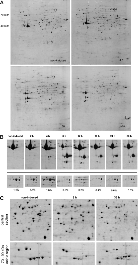
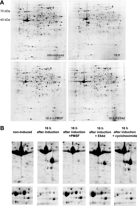
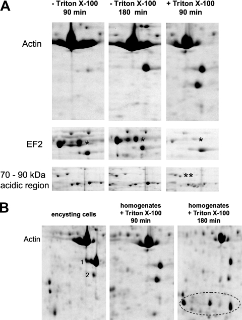
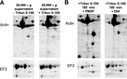
Acanthamoeba castellanii is a facultative pathogen that has a two-stage life cycle comprising the vegetatively growing trophozoite stage and the dormant cyst stage. Cysts are formed when the cell encounters unfavorable conditions, such as environmental stress or food deprivation. Due to their rigid double-layered wall, Acanthamoeba cysts are highly resistant to antiamoebic drugs. This is problematic as cysts can survive initially successful chemotherapeutic treatment and cause relapse of the disease. We studied the Acanthamoeba encystment process by using two-dimensional gel electrophoresis (2DE) and found that most changes in the protein content occur early in the process. Truncated actin isoforms were found to abound in the encysting cell, and the levels of translation elongation factor 2 (EF2) were sharply decreased, indicating that the rate of protein synthesis must be low at this stage. In the advanced stage of encystment, however, EF2 levels and the trophozoite proteome were partly restored. The protease inhibitors PMSF (phenylmethylsulfonyl fluoride) and E64d [(2S,3S)-trans-epoxysuccinyl-L-leucylamido-3-methylbutane ethyl ester] inhibited the onset of encystment, whereas the protein synthesis inhibitor cycloheximide was ineffective. Changes in the protein profile, similar to those of encysting cells, could be observed with trophozoite homogenates incubated at room temperature for several hours. Interestingly, these changes could be inhibited significantly by cysteine protease inhibitors but not by inhibitors against other proteases. Taken together, we conclude that the encystment process in A. castellanii is of a bipartite nature consisting of an initial phase of autolysis and protein degradation and an advanced stage of restoration accompanied by the expression of encystment-specific genes.


Figures

Fig. 1.
Fig. 1.
Course of encystment in A. castellanii PAT06. (A) The cells were induced to encyst in starvation medium and harvested at the time points given. Subsequently, samples were prepared for 2DE. Spot 1, actin isoform; spot 2, another actin isoform; spot 3, actin; spot 4, translation elongation factor 2 (EF2); spot 5, unidentified encystment specific protein. The 2D gels (12.5% polyacrylamide) have a pI range of 3 (left) to 10 (right). The molecular masses are indicated next to the top left gel. (B) Sections from 2DE gels, including more time points. The numbers refer to the same spots or proteins as in panel A. The proportion of EF2 of the total protein visualized on the 2D gels is given below the respective images. (C) Two sections from 2D gels corresponding to noninduced cells and cells 8 h and 36 h after induction of encystment. In the 70- to 90-kDa region in the acidic part of the gel, several trophozoite proteins are marked by asterisks; these proteins disappear 8 h after induction and reappear later in encystment.
Fig. 2.
Fig. 2.
The protease inhibitor PMSF prevents cells from starting the encystment process. (A) The cells were placed in encystment medium either without further additive or with the protease inhibitor PMSF (1 mM) or E64d (100 μM). The numbers refer to the same spots or proteins as in Fig. 1. The 2D gels (12.5% polyacrylamide) have a pI range of 3 (left) to 10 (right). The molecular masses are indicated next to the top left gel. (B) Close-up of actin and EF2 in noninduced cells, encysting cells without further additive, and encysting cells with either PMSF (1 mM), E64d (100 μM), or the protein synthesis inhibitor cycloheximide (100 μM). The numbers refer to the same spots or proteins as in Fig. 1.
Fig. 3.
Fig. 3.
The protein degradation observed in encysting cells also occurs in homogenates of noninduced trophozoites. (A) Noninduced trophozoites were lysed in a Dounce homogenizer, and the resulting homogenates were incubated at RT under shaking for various periods of time either without any additive or with 0.1% Triton X-100. The asterisks in the acidic, high-molecular-mass region on the gel mark the same proteins as in Fig. 1C. (B) After extended incubation at RT (180 min) with 0.1% Triton X-100, protein fragments that are not observed in encysting cells become apparent (circled).
Fig. 4.
Fig. 4.
The proteolytic activity observed in trophozoite homogenates resides in the large organellar fraction and can be inhibited with E64. (A) After homogenization, the large organelle fraction was removed from the trophozoite homogenates by centrifugation, and supernatants were incubated at room temperature. Translation elongation factor 2 is marked by an asterisk in panels A and B. (B) The proteolytic activity in trophozoite homogenates can be inhibited only by E64 (30 μM), not by PMSF (3 mM).

Similar articles

Cited by

References

    1. Bouyer S., Rodier M. H., Guillot A., Héchard Y. 2009. Acanthamoeba castellanii: proteins involved in actin dynamics, glycolysis, and proteolysis are regulated during encystation. Exp. Parasitol. 123:90–94 - PubMed
    1. Bowers B., Korn E. D. 1969. The fine structure of Acanthamoeba castellanii (Neff strain). II. Encystment. J. Cell Biol. 41:786–805 - PMC - PubMed
    1. Chen L., Orfeo T., Gilmartin G., Bateman E. 2004. Mechanism of cyst specific protein 21 mRNA induction during Acanthamoeba differentiation. Biochim. Biophys. Acta 1691:23–31 - PubMed
    1. De Jonckheere J. F. 1991. Ecology of Acanthamoeba. Rev. Infect. Dis. 13(Suppl. 5):S385–S387 - PubMed
    1. Dudley R., Alsam S., Khan N. A. 2008. The role of proteases in the differentiation of Acanthamoeba castellanii. FEMS Microbiol. Lett. 286:9–15 - PubMed

Publication types

MeSH terms

LinkOut - more resources