. 2010 May;76(9):3004-14.
doi: 10.1128/AEM.02085-09. Epub 2010 Feb 26.

Identification of a dehydrogenase required for lactose metabolism in Caulobacter crescentus

Affiliations

Identification of a dehydrogenase required for lactose metabolism in Caulobacter crescentus

Benjamin H Arellano et al. Appl Environ Microbiol. 2010 May.

Abstract

Caulobacter crescentus, which thrives in freshwater environments with low nutrient levels, serves as a model system for studying bacterial cell cycle regulation and organelle development. We examined its ability to utilize lactose (i) to gain insight into the metabolic capacities of oligotrophic bacteria and (ii) to obtain an additional genetic tool for studying this model organism, aiming to eliminate the basal enzymatic activity that hydrolyzes the chromogenic substrate 5-bromo-4-chloro-3-indolyl-beta-d-galactopyranoside (X-gal). Using a previously isolated transposon mutant, we identified a gene, lacA, that is required for growth on lactose as the sole carbon source and for turning colonies blue in the presence of X-gal. LacA, which contains a glucose-methanol-choline (GMC) oxidoreductase domain, has homology to the flavin subunit of Pectobacterium cypripedii's gluconate dehydrogenase. Sequence comparisons indicated that two genes near lacA, lacB and lacC, encode the other subunits of the membrane-bound dehydrogenase. In addition to lactose, all three lac genes are involved in the catabolism of three other beta-galactosides (lactulose, lactitol, and methyl-beta-d-galactoside) and two glucosides (salicin and trehalose). Dehydrogenase assays confirmed that the lac gene products oxidize lactose, salicin, and trehalose. This enzymatic activity is inducible, and increased lac expression in the presence of lactose and salicin likely contributes to the induction. Expression of lacA also depends on the presence of the lac genes, implying that the dehydrogenase participates in induction. The involvement of a dehydrogenase suggests that degradation of lactose and other sugars in C. crescentus may resemble a proposed pathway in Agrobacterium tumefaciens.


Figures
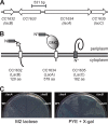
FIG. 1.
FIG. 1.
lacA, lacB, and lacC; their predicted protein products; and plate phenotypes of deletion mutants. (A) CC1632 (lacB), CC1634 (lacA), and CC1635 (lacC) encode three subunits of a dehydrogenase that is involved in lactose metabolism in C. crescentus. Arrows indicate the lengths and orientations of open reading frames. CC1633 encodes a hypothetical protein (45). The bar above CC1633 and CC1634 shows the 531-bp fragment (PlacA) used in the pJC404 reporter construct for examining expression. (B) Sequence analysis using MEMSAT3 (28) suggested that LacA, LacB, and LacC associate with the cell membrane according to the topologies depicted. All three proteins contain signal peptides (indicated by gray bars) at the amino termini (N). The leader peptides of LacA and LacC resemble those used for export via the twin-arginine translocation pathway because they each include a motif of two arginines (*) (13). LacA and LacB are predicted to have additional transmembrane segments (illustrated by white bars). LacB, a cytochrome c family protein, contains a CXXCH heme-binding motif (represented by a star), while LacA contains a GMC oxidoreductase domain (represented by a shaded ellipse) (19, 39). (C) As opposed to wild-type cells, lac deletion mutants did not form colonies on M2 minimal media with lactose as the sole carbon source (left panel). On PYE rich media with X-gal, wild-type and ΔlacB colonies turned blue, whereas ΔlacA and ΔlacC colonies stayed white (right panel). ΔlacB colonies were generally less blue than were the wild-type ones.
FIG. 2.
FIG. 2.
Comparison of wild-type and ΔlacA strains on agar media containing X-gal. Wild-type and mutant strains carrying the empty vector, mutants carrying a plasmid with lacA, and mutants carrying a plasmid with E. coli lacZ (lacZEc) were grown on M2 minimal media plus 0.2% lactose, PYE media plus tetracycline, or PYE alone, as indicated below each panel. The far right panel [PYE (zoom)] signposts the strain genotype in each quadrant of all panels and shows the close-up of colonies grown on PYE media with X-gal. Mutant strains with the empty vector or lacZEc failed to grow on M2 plus lactose. The plasmid vector (pCM62) and its derivatives (pJC327, pJC389) all confer resistance to tetracycline. Loss of the plasmid carrying lacA or E. coli lacZ from ΔlacA strains can be detected visually as growth of white or partially white colonies on PYE media.
FIG. 3.
FIG. 3.
ONPG hydrolysis activity in wild-type and ΔlacA strains. (A) Wild-type (lacA+) cells induced with lactose showed the highest level of ONPG hydrolysis, compared with uninduced wild-type cells and induced or uninduced ΔlacA cells. (B) ONPG hydrolysis by induced lacA+ cells and uninduced ΔlacA cells was assessed in the absence or presence of different inhibitors, as follows: sodium azide (azide), chloramphenicol (Cm), or SDS and chloroform (SDS+CHCl3). Levels of ONPG hydrolysis were measured as described in Materials and Methods and normalized according to the average activity in induced lacA+ cells, without metabolic inhibitor. Error bars indicate standard deviations.
FIG. 4.
FIG. 4.
lac genes required for utilization of specific carbon sources (A) Phenotype MicroArray analysis indicated that the lacA deletion inhibited growth on six carbon sources, the chemical structures of which are depicted. Salicin is a β-glucoside, and trehalose is an α-glucoside, while lactose, lactulose, lactitol, and methyl-β-d-galactoside are all β-galactosides. (B) Deletion of any lac gene prevented colony formation on M2 agar with salicin in the NA1000 background (top panel) but reduced growth only in the CB15 background (middle panel). The bottom panel indicates the genotype in each sector of the plate. For the other five sugars, deletion of any lac gene prevented growth in both strain backgrounds.
FIG. 5.
FIG. 5.
Dehydrogenase activity, as measured by reduction of DCIP, in wild-type (lacA+) and ΔlacA mutant strains under different conditions. (A) Dehydrogenase activities of wild-type and mutant cells, grown in the absence or presence of the lactose inducer (uninduced or induced), were assessed with xylose, lactose, or salicin as the electron donor. Induced lacA+ cells incubated with lactose or salicin exhibited the highest levels of activity. (B) A plasmid carrying the lacA gene complements the deficiency in dehydrogenase activity in the ΔlacA mutant. Wild-type and mutant cells with the complementing lacA+ plasmid (pJC389) or the empty vector (pCM62) were grown in the absence or presence of lactose (uninduced or induced) and then analyzed with lactose as the electron donor. Dehydrogenase activities were measured as described in Materials and Methods, with 1 unit of activity defined as the change in OD600 per minute per unit of cell density. Reactions without cells showed no activity. Error bars indicate standard deviations.
FIG. 6.
FIG. 6.
Variations in lacA expression in different growth media, as determined by β-galactosidase assays using a PlacA-lacZEc reporter fusion. (A) Wild-type cells carrying the reporter construct (pJC404) were grown in PYE broth, M2 minimal media plus 0.2% glucose (M2G), or M2 plus 0.2% xylose (M2X), in the absence or presence of 0.2% lactose as the inducer; cells grown to mid-log phase were harvested to measure levels of β-galactosidase activity. (B) Wild-type (lacA+) and ΔlacA cells were grown in M2G with no other sugar, 0.2% xylose, 0.2% lactose, or 0.02% salicin and harvested for β-galactosidase assays. Error bars indicate standard deviations.

Similar articles

Cited by

References

    1. Ausubel, F. M., R. Brent, R. E. Kingston, D. D. Moore, J. G. Seidman, J. A. Smith, and K. Struhl (ed.). 1998. Current protocols in molecular biology. John Wiley & Sons, New York, NY.
    1. Baldrian, P., and V. Valaskova. 2008. Degradation of cellulose by basidiomycetous fungi. FEMS Microbiol. Rev. 32:501-521. - PubMed
    1. Bastedo, D. P., and G. T. Marczynski. 2009. CtrA response regulator binding to the Caulobacter chromosome replication origin is required during nutrient and antibiotic stress as well as during cell cycle progression. Mol. Microbiol. 72:139-154. - PubMed
    1. Blondelet-Rouault, M. H., J. Weiser, A. Lebrihi, P. Branny, and J. L. Pernodet. 1997. Antibiotic resistance gene cassettes derived from the omega interposon for use in E. coli and Streptomyces. Gene 190:315-317. - PubMed
    1. Bochner, B. R., P. Gadzinski, and E. Panomitros. 2001. Phenotype microarrays for high-throughput phenotypic testing and assay of gene function. Genome Res. 11:1246-1255. - PMC - PubMed

Publication types

LinkOut - more resources