. 2010 Jun;298(6):H1652-60.
doi: 10.1152/ajpheart.01128.2009. Epub 2010 Feb 26.

Heterogeneous ventricular sympathetic innervation, altered beta-adrenergic receptor expression, and rhythm instability in mice lacking the p75 neurotrophin receptor

Affiliations

Heterogeneous ventricular sympathetic innervation, altered beta-adrenergic receptor expression, and rhythm instability in mice lacking the p75 neurotrophin receptor

Christina U Lorentz et al. Am J Physiol Heart Circ Physiol. 2010 Jun.

Abstract

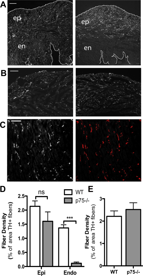
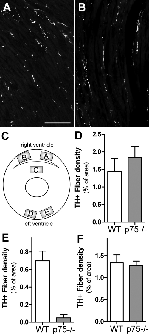
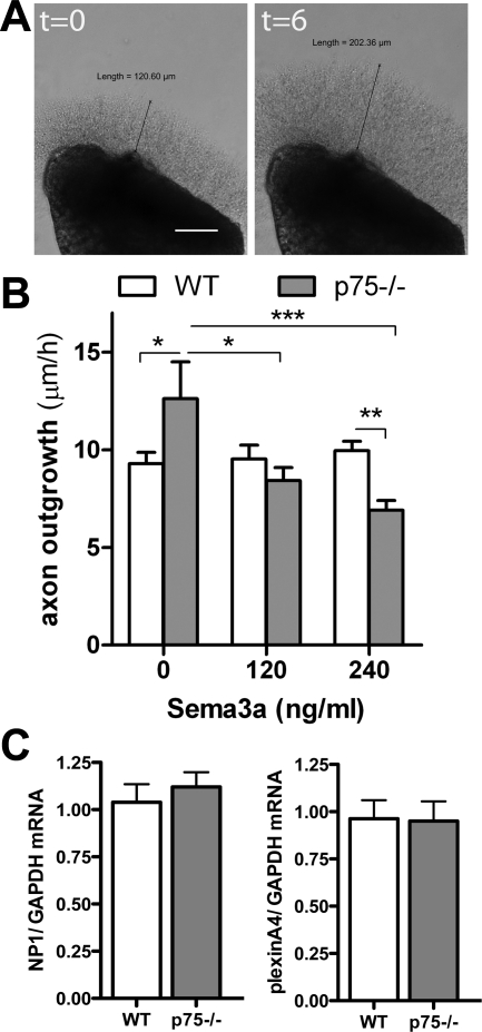
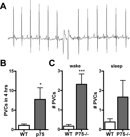
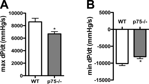
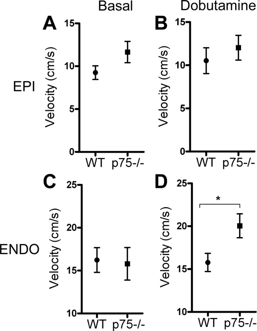
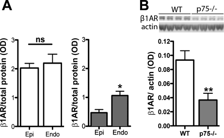
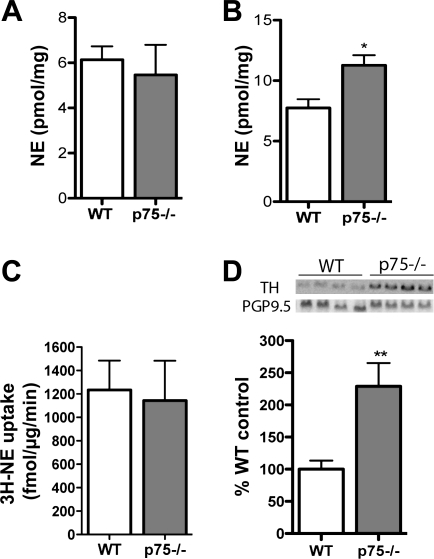
Sympathetic nerves stimulate cardiac function through the release of norepinephrine and the activation of cardiac beta(1)-adrenergic receptors. The sympathetic innervation of the heart is sculpted during development by chemoattractive factors including nerve growth factor (NGF) and the chemorepulsive factor semaphorin 3a. NGF acts through the TrkA receptor and the p75 neurotrophin receptor (p75(NTR)) in sympathetic neurons. NGF stimulates sympathetic axon extension into the heart through TrkA, but p75(NTR) modulates multiple coreceptors that can either stimulate or inhibit axon outgrowth. In mice lacking p75(NTR), the sympathetic innervation density in target tissues ranges from denervation to hyperinnervation. Recent studies have revealed significant changes in the sympathetic innervation density of p75NTR-deficient (p75(NTR-/-)) atria between early postnatal development and adulthood. We examined the innervation of adult p75(NTR-/-) ventricles and discovered that the subendocardium of the p75(NTR-/-) left ventricle was essentially devoid of sympathetic nerve fibers, whereas the innervation density of the subepicardium was normal. This phenotype is similar to that seen in mice overexpressing semaphorin 3a, and we found that sympathetic axons lacking p75(NTR) are more sensitive to semaphorin 3a in vitro than control neurons. The lack of subendocardial innervation was associated with decreased dP/dt, altered cardiac beta(1)-adrenergic receptor expression and sensitivity, and a significant increase in spontaneous ventricular arrhythmias. The lack of p75(NTR) also resulted in increased tyrosine hydroxylase content in cardiac sympathetic neurons and elevated norepinephrine in the right ventricle, where innervation density was normal.


Figures

Fig. 1.
Fig. 1.
Fiber distribution is altered in the p75 neurotrophin receptor-deficient (p75NTR−/−) left ventricle (LV). Sympathetic fiber distribution was identified by tyrosine hydroxylase (TH) immunoreactivity. A: representative pictures taken at ×5 magnification showing the entire width of the LV free wall revealing the altered pattern of sympathetic innervation in p75NTR−/− mice (right) compared with wild-type (WT) mice (left). This low magnification was not used for quantitative analysis. The edges of the tissue have been outlined for clarity. Scale bar = 0.25 mm. B: sympathetic fiber distribution in the right ventricle (RV) of WT mice (left) and p75NTR−/− mice (right). Scale bar = 0.1 mm. C: example of the threshold discrimination used to determine innervation density in which the TH-positive nerves above the threshold are highlighted. Innervation density was then expressed as the percent area that was above the determined threshold (TH+ fibers). Scale bar = 0.05 mm. D: quantification of sympathetic fiber density in the subendocardium (en and Endo) and subepicardium (ep and Epi) of WT and p75NTR−/− mice. ns, Not significant. Values are means ± SE; n = 4. ***P < 0.001. E: quantification of sympathetic fiber density of the RV in WT and p75NTR−/− mice. Values are means ± SE; n = 4.
Fig. 2.
Fig. 2.
Innervation density is normal in the p75NTR−/− subepicardium. A and B: representative pictures at ×20 magnification of TH+ fibers in the WT (A) and p75NTR−/− (B) subepicardium that were used for quantitative analysis. Scale bar = 0.1 mm. C: diagram showing the regions (areas A–E) analyzed to further characterize the innervation density of WT and p75NTR−/− ventricles. D–F: quantification of sympathetic fiber density in the WT and p75NTR−/− RV (D), interventricular septum (E), and LV (F). Values are means ± SE; n = 7.
Fig. 3.
Fig. 3.
p75NTR−/− sympathetic axons are more sensitive to semaphorin 3a (Sema3a) growth cone collapse/inhibition. A: Representative stellate ganglia explants at time 0 (left) and 6 h later (right). Scale bar = 0.1 mm. B: quantification of axon growth rate from WT and p75NTR−/− sympathetic ganglia in the presence of 0, 120, or 240 ng/ml Sema3a. Values are means ± SE; n = 3. *P < 0.05; **P < 0.01; ***P < 0.001. C: neuropilin (NP)-1 (left) and plexin A4 (right) mRNA normalized to GAPDH in WT and p75NTR−/− stellate ganglia. Values are means ± SE; n = 5.
Fig. 4.
Fig. 4.
Mice lacking p75NTR have increased spontaneous ventricular arrhythmias. A: representative ECG showing spontaneous premature ventricular complexes (PVCs) in a p75NTR−/− mouse. B: p75NTR−/− mice experienced more spontaneous PVCs than WT mice. Values are means ± SE; n = 8–9. *P < 0.05. C: number of PVCs in WT and p75NTR−/− mice during 2 h of the awake phase and sleep phase. Values are means ± SE; n = 8–9. ***P < 0.01.
Fig. 5.
Fig. 5.
dP/dtmax and dP/dtmin are low in p75NTR−/− mice. A: baseline dP/dtmax in WT and p75NTR−/− mice. B: dP/dtmin in WT and p75NTR−/− mice. Values are means ± SE; n = 6–7. *P < 0.05.
Fig. 6.
Fig. 6.
p75NTR−/− mice have an enhanced response to β1-adrenergic receptor (β1AR) stimulation in the denervated subendocardium. A and B: peak radial thickening velocity of the subepicardium at early diastole in WT and p75NTR−/− mice as determined by tissue Doppler analysis before (A) and after (B) an intraperitoneal injection of dobutamine (32 μg/kg). C and D: peak radial thickening velocity of the subendocardium in WT and p75NTR−/− mice determined at early diastole before (C) or after (D) an intraperitoneal injection of dobutamine (32 μg/kg). Values are means ± SE; n = 4. *P < 0.05.
Fig. 7.
Fig. 7.
β1AR expression is low, and its distribution is altered in the p75NTR−/− LV. A: β1AR protein in the subendocardium and subepicardium of WT (left; open bars) and p75NTR−/− mice (right; shaded bars) normalized to total protein. Values are means ± SE; n = 4. *P < 0.05. B: representative Western blot (top) and quantification (bottom) of β1ARs and actin in WT and p75NTR−/− LVs. Values are means ± SE; n = 6. *P < 0.05; **P < 0.01.
Fig. 8.
Fig. 8.
Norephinephrine (NE) content, NE uptake, and TH levels in the LV. A: NE content in the p75NTR−/− LV was not significantly different than that in the WT LV. Values are means ± SE; n = 5–6. B: NE is elevated in the RV of p75NTR−/− mice. Values are means ± SE; n = 5–6. *P < 0.05. C: NE uptake in p75−/− mouse ventricles is identical to uptake in WT ventricles. Values are means ± SE; n = 6. D: TH protein, normalized to the pan-neuronal marker protein gene product 9.5 (PGP9.5), is elevated in the LV of p75NTR−/− mice compared with WT mice. Values are means ± SE; n = 5–6. **P < 0.01.

Comment in

References

    1. Aronow WS, Ahn C, Mercando AD, Epstein S. Circadian variation of sudden cardiac death or fatal myocardial infarction is abolished by propranolol in patients with heart disease and complex ventricular arrhythmias. Am J Cardiol 74: 819–821, 1994 - PubMed
    1. Bandtlow C, Dechant G. From cell death to neuronal regeneration, effects of the p75 neurotrophin receptor depend on interactions with partner subunits. Sci STKE 2004: pe24, 2004 - PubMed
    1. Ben-Zvi A, Ben-Gigi L, Klein H, Behar O. Modulation of semaphorin3A activity by p75 neurotrophin receptor influences peripheral axon patterning. J Neurosci 27: 13000–13011, 2007 - PMC - PubMed
    1. Bjerre B, Bjorklund A, Mobley W, Rosengren E. Short- and long-term effects of nerve growth factor on the sympathetic nervous system in the adult mouse. Brain Res 94: 263–277, 1975 - PubMed
    1. Brunet S, Aimond F, Li H, Guo W, Eldstrom J, Fedida D, Yamada KA, Nerbonne JM. Heterogeneous expression of repolarizing, voltage-gated K+ currents in adult mouse ventricles. J Physiol 559: 103–120, 2004 - PMC - PubMed

Publication types

MeSH terms