. 2010 Apr 1;184(7):3357-66.
doi: 10.4049/jimmunol.0902398. Epub 2010 Feb 26.

MHC class I molecules with Superenhanced CD8 binding properties bypass the requirement for cognate TCR recognition and nonspecifically activate CTLs

Affiliations

MHC class I molecules with Superenhanced CD8 binding properties bypass the requirement for cognate TCR recognition and nonspecifically activate CTLs

Linda Wooldridge et al. J Immunol. .

Abstract

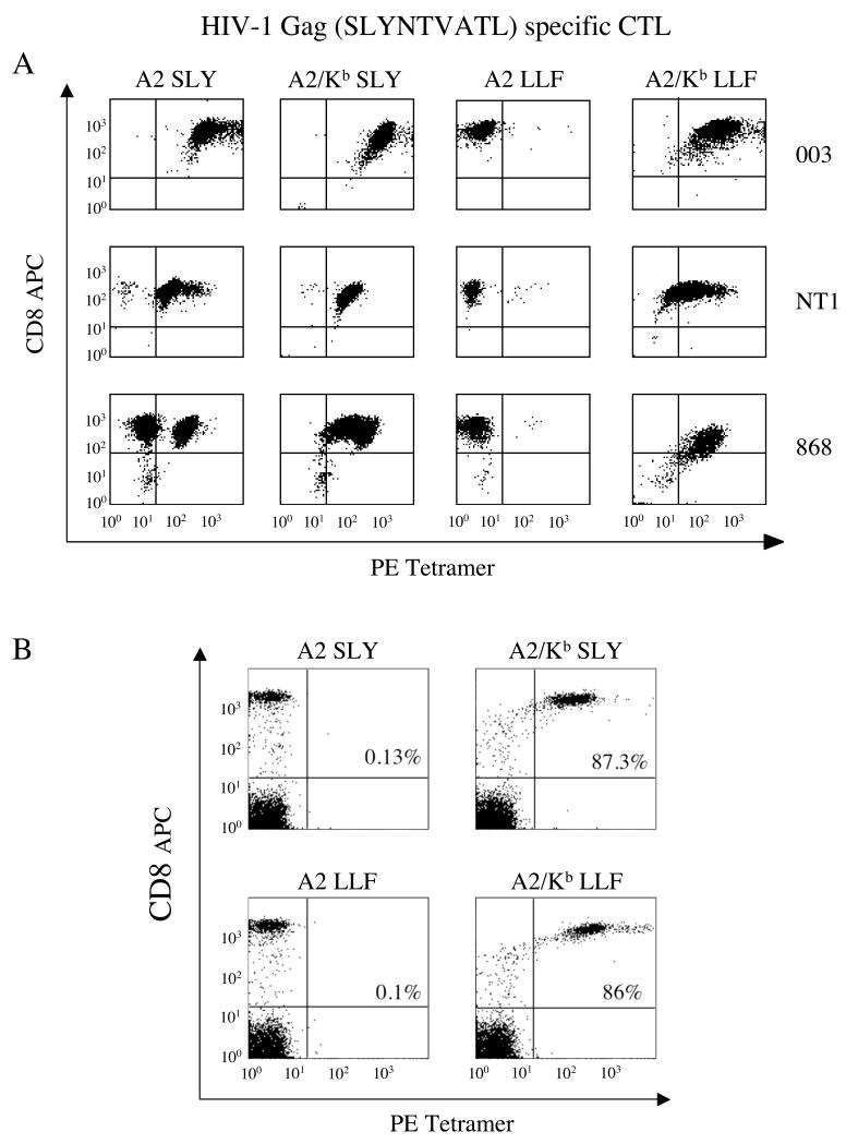
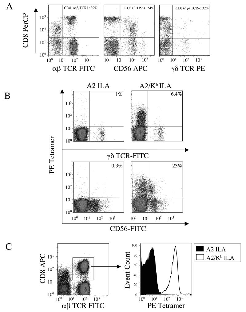
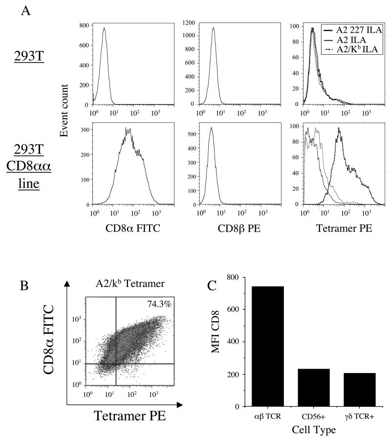
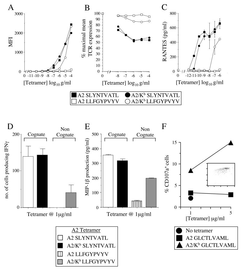
CD8(+) CTLs are essential for effective immune defense against intracellular microbes and neoplasia. CTLs recognize short peptide fragments presented in association with MHC class I (MHCI) molecules on the surface of infected or dysregulated cells. Ag recognition involves the binding of both TCR and CD8 coreceptor to a single ligand (peptide MHCI [pMHCI]). The TCR/pMHCI interaction confers Ag specificity, whereas the pMHCI/CD8 interaction mediates enhanced sensitivity to Ag. Striking biophysical differences exist between the TCR/pMHCI and pMHCI/CD8 interactions; indeed, the pMHCI/CD8 interaction can be >100-fold weaker than the cognate TCR/pMHCI interaction. In this study, we show that increasing the strength of the pMHCI/CD8 interaction by approximately 15-fold results in nonspecific, cognate Ag-independent pMHCI tetramer binding at the cell surface. Furthermore, pMHCI molecules with superenhanced affinity for CD8 activate CTLs in the absence of a specific TCR/pMHCI interaction to elicit a full range of effector functions, including cytokine/chemokine release, degranulation and proliferation. Thus, the low solution binding affinity of the pMHCI/CD8 interaction is essential for the maintenance of CTL Ag specificity.


Figures

Figure 1
Figure 1. The exquisite specificity of pMHCI tetramer staining is lost when the strength of the pMHCI/CD8 interaction is increased by ~15-fold
A. The 003 or NT1 CTL clones (105 cells) or the 868 CTL line (2.5×10⁵ cells), all specific for HIV-1 p17 Gag77-85, were stained with 1μg of the PE-conjugated tetramers A2 SLYNTVATL, A2/Kb SLYNTVATL, A2 LLFGYPVYV or A2/Kb LLFGYPVYV in 20μl PBS for 20 minutes at 37°C. Cells were then stained with APC-conjugated anti-CD8 and 7-AAD for 30 minutes on ice, washed twice and resuspended in PBS. Data were acquired using a FACSCalibur flow cytometer and analyzed with CellQuest software. B. 2.5×10⁵ PBMC were suspended in 250 μl FACS buffer (2% FCS/PBS), then stained with 1μg of the PE-conjugated tetramers A2 SLYNTVATL, A2/Kb SLYNTVATL, A2 LLFGYPVYV or A2/Kb LLFGYPVYV for 20 minutes at 37°C. Each sample was subsequently stained with APC-conjugated anti-CD8, PerCP-conjugated anti-CD3 and 7-AAD for 30 minutes on ice, washed twice and resuspended in FACS buffer. Data were acquired using a FACSCalibur flow cytometer and analysed with CellQuest software by gating on the live CD3+ population. The values shown represent the percent of CD3+CD8+ cells that stain with the indicated tetramer.
Figure 2
Figure 2. A2/Kb tetramers bind the majority of CTL in peripheral blood
A. 2.5×10⁵ PBMC from an A2+ donor were stained with PerCP-conjugated anti-CD8, 7-AAD and either FITC-conjugated anti-αβ-TCR, APC-conjugated anti-CD56 or PE-conjugated anti-γδ-TCR for 30 minutes on ice, washed twice and resuspended in PBS. B. 2.5×10⁵ A2+PBMC were stained with 10μg/ml of the PE-conjugated tetramers A2 ILAKFLHWL or A2/Kb ILAKFLHWL for 20 minutes at 37°C. After washing, cells were subsequently stained with 7-AAD and either FITC-conjugated anti-γδ-TCR or FITC-conjugated anti-CD56 for 30 minutes on ice, washed twice and resuspended in PBS. C. 2.5×10⁵ A2+ PBMC were stained with 10μg/ml of the PE-conjugated tetramers A2 ILAKFLHWL or A2/Kb ILAKFLHWL for 20 minutes at 37°C. After washing, cells were stained with APC-conjugated anti-CD8, FITC-conjugated anti-αβ-TCR and 7-AAD for 30 minutes on ice, washed twice and resuspended in PBS. In A, B and C, data were acquired using a FACSCalibur flow cytometer and analyzed with FlowJo software.
Figure 3
Figure 3. Non-specific A2/Kb tetramer binding is influenced by CD8 cell surface density
A and B. 2×10⁵ 293T cells were incubated +/− 10 μg/ml of the PE-conjugated tetramers A2 D227K/T228A ILAKFLHWL, A2 ILAKFLHWL or A2/Kb ILAKFLHWL for 20 minutes at 37°C, then stained with 7-AAD and either FITC-conjugated anti-CD8 or PE-conjugated anti-CD8β for 30 minutes on ice, washed twice and resuspended in PBS. C. 2.5×10⁵ PBMC were stained with PerCP-conjugated anti-CD8, 7-AAD and either FITC-conjugated anti-αβ-TCR, APC-conjugated anti-CD56 or PE-conjugated anti-γδ-TCR for 30 minutes on ice, washed twice and resuspended in PBS. In A, B and C, data were acquired using a FACSCalibur flow cytometer and analyzed with FlowJo software.
Figure 4
Figure 4. A2/Kb tetramers can activate CTL in the absence of a specific TCR/pMHCI interaction
A. 105 003 CTL were suspended in 20μl PBS and stained with the PE-conjugated tetramers A2 SLYNTVATL, A2/Kb SLYNTVATL, A2 LLFGYPVYV or A2/Kb LLFGYPVYV at the indicated concentrations and 7-AAD for 20 minutes at 37°C. Cells were then washed twice and resuspended in PBS. Data were acquired using a FACSCalibur flow cytometer and analyzed with CellQuest software. B. 105 003 CTL were suspended in 40μl R2 with the PE-conjugated tetramers A2 SLYNTVATL, A2/Kb SLYNTVATL, A2 LLFGYPVYV or A2/Kb LLFGYPVYV at the indicated concentrations for 30 minutes at 37°C. Cells were subsequently stained with FITC-conjugated anti-αβ-TCR, 7-AAD and APC-conjugated anti-CD8 for 30 minutes on ice in azide buffer (0.1% azide/2% FCS/PBS). After two washes, data were acquired using a FACSCalibur flow cytometer and analyzed with CellQuest software. C. 5×10⁵ 003 CTL were incubated with the PE-conjugated tetramers A2 SLYNTVATL, A2/Kb SLYNTVATL, A2 LLFGYPVYV or A2/Kb LLFGYPVYV at the indicated concentrations. After 4 hours at 37°C, supernatants were harvested and assayed for RANTES, IFNγ and MIP-1β content by ELISA (only RANTES shown). D. 2×10³ 868 CTL were incubated for 4 hours at 37°C with 1μg/ml of the PE-conjugated tetramers A2 SLYNTVATL, A2/Kb SLYNTVATL, A2 LLFGYPVYV or A2/Kb LLFGYPVYV in an IFNγ ELISpot assay. E. 1.25×10⁵ 868 CTL were incubated with 1μg/ml of the PE-conjugated tetramers A2 SLYNTVATL, A2/Kb SLYNTVATL, A2 LLFGYPVYV or A2/Kb LLFGYPVYV for 4 hours at 37°C. The supernatant was subsequently assayed for MIP-1β content by ELISA. Figures (C-E) show the mean ± SD of two replicate assays. Results similar to (A-E) were also obtained with tetramers conjugated to fluorochromes other than PE (data not shown). F. 003 CTL were incubated with the PE-conjugated tetramers A2 SLYNTVATL, A2/Kb SLYNTVATL, A2 GLCTLVAML or A2/KbGLCTLVAML at the indicated concentrations for 4 hours at 37°C, then stained with APC-conjugated anti-CD8 for 20 minutes on ice and assayed for CD107a mobilization as described in the Materials and Methods. The inset plot shows staining for APC-conjugated anti-CD8 on the x-axis and PE-conjugated A2/Kb GLCTLVAML tetramer (5μg/ml) on the y-axis. Back-gated tetramer+CD107a+ cells are shown in black and tetramer+CD107a cells are shown in grey. TetramerhighCD8high cells are preferentially activated by the A2/Kb tetramer.
Figure 5
Figure 5. Cell surface-expressed A2/Kb activates CTL in the absence of cognate antigen
A. 2.5×10⁴ Mel13, 003 or ILA1 CTL were incubated for 12 hours at 37°C with 105 C1R cells stably transfected to express equal levels of either A2 or A2/Kb at the cell surface. Supernatant was subsequently assayed for MIP-1β content by ELISA. The mean ± SD of two replicate assays is shown. B. 2.5×10⁴ Mel13 CTL were incubated for 12 hours at 37°C with 105 C1R cells stably transfected to express either A2 or A2/Kb at the cell surfaceSupernatant was assayed for IFNγ and TNFα content by cytokine bead array. C. CD107a expression by ILA1 and Mel13 CTL following a 12 hour incubation at 37°C with C1R cells stably transfected to express either A2 or A2/Kb on the cell surface. For (A-C), C1R cells were not previously pulsed with peptide.
Figure 6
Figure 6. Cell surface-expressed A2/Kb primes non-specific expansion of CD8+ cells
106 A2+ PBMC were incubated with 2×10⁵ irradiated A2 D227K/T228A, A2 or A2/Kb C1R cells that had previously been pulsed with 1μM ELAGIGILTV (Melan-A26-35) peptide in R10. From day 3, IL-2 was added in increments to reach a maximum concentration of 200 IU/ml by day 10. Lines were subsequently stained with PE-conjugated A2 ELAGIGILTV tetramer followed by APC-conjugated anti-CD8 and 7-AAD. Data were acquired using a FACSCalibur flow cytometer and analyzed with FlowJo software.
Figure 7
Figure 7. Non-cognate A2/Kb-mediated CTL activation and tetramer binding is not influenced by MHCI restriction
A. 2.5×10⁵ PBMC were suspended in 250μl FACS buffer (2% FCS/PBS) and stained with FITC-conjugated anti-A2 and 7-AAD for 30 minutes on ice, then washed twice and resuspended in PBS. For pMHCI tetramer staining experiments, 2.5×10⁵ PBMC were suspended in 50μl FACS buffer (2% FCS/PBS) and incubated +/− 10μg/ml unconjugated anti-CD8 for 20 minutes on ice, then stained with 10μg/ml of the PE-conjugated tetramers A2 ILAKFLHWL or A2/Kb ILAKFLHWL for 45 minutes on ice. After washing, cells were subsequently stained with APC-conjugated anti-CD8 and 7-AAD, washed again and resuspended in PBS. Data were acquired using a FACSCalibur flow cytometer and analyzed with FlowJo software. B. 2.5×10⁴ CTL were incubated for 12 hours at 37°C with 105 unpulsed C1R cells expressing either A2 or A2/Kb on the cell surface. The following CTL clones were used: (i) the HLA A*6801-restricted CTL clone c23, specific for the HIV-1 Tat-derived epitope ITKGLGISYGR (residues 38-48); (ii) the HLA B*0702-restricted CTL clone KD4, specific for the EBV EBNA3A-derived epitope RPPIFIRRL (residues 379-387); (iii) the HLA B*0801-restricted CTL clone LC13, specific for the EBV EBNA3A-derived epitope FLRGRAYGL (residues 339-347); and, (iv) the HLA B*3508-restricted CTL clone SB27, specific for the EBV BZLF1-derived epitope LPEPLPQGQLTAY (residues 52-64). Supernatant was subsequently assayed for MIP-1β content by ELISA. The mean ± SD of two replicate assays is shown.

Similar articles

Cited by

References

    1. Davis MM, Boniface JJ, Reich Z, Lyons D, Hampl J, Arden B, Chien Y. Ligand recognition by alpha beta T cell receptors. Annu Rev Immunol. 1998;16:523–544. - PubMed
    1. Rudolph MG, Wilson IA. The specificity of TCR/pMHC interaction. Curr Opin Immunol. 2002;14:52–65. - PubMed
    1. Zamoyska R. CD4 and CD8: modulators of T-cell receptor recognition of antigen and of immune responses? Curr Opin Immunol. 1998;10:82–87. - PubMed
    1. Holler PD, Kranz DM. Quantitative analysis of the contribution of TCR/pepMHC affinity and CD8 to T cell activation. Immunity. 2003;18:255–264. - PubMed
    1. Gakamsky DM, Luescher IF, Pramanik A, Kopito RB, Lemonnier F, Vogel H, Rigler R, Pecht I. CD8 kinetically promotes ligand binding to the T-cell antigen receptor. Biophys J. 2005;89:2121–2133. - PMC - PubMed

Publication types

MeSH terms

Substances