

Case Reports
doi: 10.3174/ajnr.A1873.
Epub 2010 Feb 25.
Cerebral white matter lesions may be partially reversible in patients with carotid artery stenosis
Affiliations
- PMID: 20190206
- PMCID: PMC7965479
- DOI: 10.3174/ajnr.A1873
Item in Clipboard
Case Reports
Cerebral white matter lesions may be partially reversible in patients with carotid artery stenosis
AJNR Am J Neuroradiol.
2010 Aug.
Abstract
Contrary to the common belief that age-related WMLs (also known as leukoaraiosis) are a progressive condition, a case of partial reversal of WMLs shortly after carotid artery stenting is described. A 75-year-old man presented with frequent TIAs, which were attributed to right ICA stenosis. He subsequently underwent successful carotid artery stenting. Follow-up MR imaging a week after the procedure showed improvement of WMLs in the right cerebral hemisphere. Pixel-by-pixel image analysis showed that the reversed WMLs tended to have higher lambda1 and lower signal intensity on b = 0 images compared with nonreversed lesions, but by only approximately 10%.
Figures
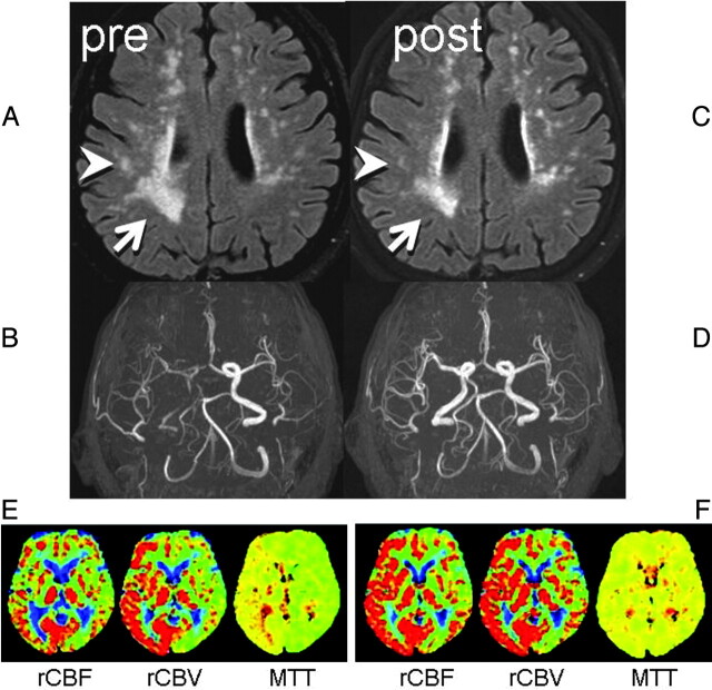
Two consecutive FLAIR images and MR angiography. A, Preprocedural FLAIR (delay time, 2200 ms; TR/TE, 8000/100 ms) demonstrates WMLs. B, Preprocedural MR angiography (TR/TE, 30/2.3 ms) shows poor visualization of the right anterior circulation branches. C, Postprocedural FLAIR a week later shows partial WML resolution in the right hemisphere (arrow/arrowhead). D, MR angiogram shows restoration of flow in the right ICA. E, Preprocedural perfusion-weighted images show MTT elongation at the right anterior circulation territory with compensatory vasodilation, indicated by increased cerebral blood volume. F, After the procedure, the MTT elongation is normalized. Increased cerebral blood flow at the same territory indicates postprocedural hyperperfusion, but the patient remained asymptomatic.
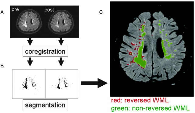
Image analysis on pre- and postprocedural images. A, Image coregistration was performed between the pre- and postprocedural FLAIR. B, Segmentation of the pixels with WMLs was then done. C, Subtraction images yielded pixels with/without reversal. Coregistration of these data onto the DWI/DTI (DWI: TR/TE, 6000/88 ms; b = 1000 s/mm2) and b = 0 imaging was performed to characterize the nature of the WMLs.
Similar articles
-
The Circle of Willis and White Matter Lesions in Patients with Carotid Atherosclerosis.J Stroke Cerebrovasc Dis. 2015 Aug;24(8):1749-54. doi: 10.1016/j.jstrokecerebrovasdis.2015.03.048. Epub 2015 Jun 19. J Stroke Cerebrovasc Dis. 2015. PMID: 26096317
-
External carotid artery angioplasty and stenting to augment cerebral perfusion in the setting of subacute symptomatic ipsilateral internal carotid artery occlusion. Case report.J Neurosurg. 2007 Dec;107(6):1217-22. doi: 10.3171/JNS-07/12/1217. J Neurosurg. 2007. PMID: 18077961
-
MR and clinical follow-up of diffusion-weighted cerebral lesions after carotid artery stenting.AJNR Am J Neuroradiol. 2005 Oct;26(9):2336-41. AJNR Am J Neuroradiol. 2005. PMID: 16219842 Free PMC article.
-
Age and Diastolic Blood Pressure Play an Important Role in the Progression of White Matter Lesions: A Meta-Analysis.Eur Neurol. 2020;83(4):351-359. doi: 10.1159/000510077. Epub 2020 Sep 9. Eur Neurol. 2020. PMID: 32906133
-
Stenting for Internal Carotid Artery Stenosis Associated with Persistent Primitive Hypoglossal Artery Using Proximal Flow Blockade and Distal Protection System: A Technical Case Report and Literature Review.J Stroke Cerebrovasc Dis. 2016 Jun;25(6):e98-e102. doi: 10.1016/j.jstrokecerebrovasdis.2016.03.026. Epub 2016 Apr 19. J Stroke Cerebrovasc Dis. 2016. PMID: 27105567 Review.
Cited by
-
Pathophysiological link between carotid atherosclerosis and cerebral white matter lesions.Sci Rep. 2025 Feb 24;15(1):6619. doi: 10.1038/s41598-025-90922-3. Sci Rep. 2025. PMID: 39994379 Free PMC article.
-
Attenuation of brain white matter lesions after lacunar stroke.Int J Prev Med. 2012 Feb;3(2):134-8. Int J Prev Med. 2012. PMID: 22347611 Free PMC article.
-
Characterizing the penumbras of white matter hyperintensities in patients with cerebral small vessel disease.Jpn J Radiol. 2023 Sep;41(9):928-937. doi: 10.1007/s11604-023-01419-w. Epub 2023 May 9. Jpn J Radiol. 2023. PMID: 37160589 Free PMC article.
-
Characterization of the Growth of Deep and Subcortical White Matter Hyperintensity on MR Imaging: A Retrospective Cohort Study.Magn Reson Med Sci. 2017 Jul 10;16(3):238-244. doi: 10.2463/mrms.mp.2016-0063. Epub 2017 Jan 13. Magn Reson Med Sci. 2017. PMID: 28090008 Free PMC article.
-
Characterizing the Penumbras of White Matter Hyperintensities and Their Associations With Cognitive Function in Patients With Subcortical Vascular Mild Cognitive Impairment.Front Neurol. 2019 Apr 12;10:348. doi: 10.3389/fneur.2019.00348. eCollection 2019. Front Neurol. 2019. PMID: 31031687 Free PMC article.
References
-
- Hachinski VC, Potter P, Merskey H. Leukoaraiosis. Arch Neurol 1987;4:21–23 - PubMed
-
- Wiszniewska M, Devuyst G, Bogousslavsky J, et al. . What is the significance of leukoaraiosis in patients with acute ischemic stroke? Arch Neurol 2000;57:967–73 - PubMed
-
- Longstreth WT, Jr, Arnold AM, Beauchamp NJ, Jr, et al. . Incidence, manifestations, and predictors of worsening white matter on serial cranial magnetic resonance imaging in the elderly. Stroke 2005;36:56–61 - PubMed
-
- Streifler JY, Eliasziw M, Benavente OR, et al. . Development and progression of leukoaraiosis in patients with brain ischemia and carotid artery disease. Stroke 2003;34:1913–16 - PubMed
-
- Brun A, Englund EA. White matter disorder in dementia of the Alzheimer type: a pathoanatomical study. Ann Neurol 1986;19:253–62 - PubMed
Publication types
MeSH terms
LinkOut - more resources
Full Text Sources
Medical
Miscellaneous