. 2010 Apr 23;285(17):12638-46.
doi: 10.1074/jbc.M109.077370. Epub 2010 Feb 27.

Drf1-dependent kinase interacts with Claspin through a conserved protein motif

Affiliations

Drf1-dependent kinase interacts with Claspin through a conserved protein motif

Daniel A Gold et al. J Biol Chem. .

Abstract

The Dbf4/Drf1-dependent kinase (DDK) is required for the initiation of DNA replication in eukaryotes. Another protein, Claspin, mediates the activation of a cellular checkpoint response to stalled replication forks and is also a regulator of replication. In this study, we found that DDK phosphorylates Claspin in vitro and forms a nuclear complex containing Cdc7, Drf1, and Claspin in Xenopus egg extracts. In addition, purified Claspin and DDK are capable of a direct in vitro interaction. We identified a conserved binding site on Claspin required for its interaction with DDK. This site corresponds to the first of two sequence repeats in the Chk1-binding domain of Claspin. Furthermore, we have established that two amino acids in this motif, Asp(861) and Gln(866), are essential for the interaction between Claspin and DDK. We found that mutant forms of Claspin incapable of interacting with DDK are still able to associate with and activate Chk1 in response to DNA replication blockages. However, Claspin-depleted egg extracts that have been reconstituted with these mutants of Claspin undergo DNA replication more slowly. These findings suggest that the interaction of DDK with Claspin mediates a checkpoint-independent function of Claspin related to DNA replication.


Figures
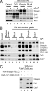
FIGURE 1.
FIGURE 1.
Claspin forms a complex with Cdc7 and Drf1 (DDK) in Xenopus egg extracts. A, IPs with control (Mock), anti-Drf1, and anti-Claspin antibodies were prepared from nuclei-free interphase egg extracts lacking or containing (dA)70-(dT)70 (pA-pT) and immunoblotted for Claspin, Drf1, Cdc7, and Plx1. Lanes 1 and 2 depict 2% of pre-IP extracts. B, interphase extracts containing sperm nuclei (4,000/μl) were incubated in the absence or presence of aphidicolin (APH). Nuclear lysates were prepared and immunoprecipitated with control (Mock), anti-Drf1, anti-Claspin, and anti-Cdc7 antibodies. These immunoprecipitates were immunoblotted for Scc2, Claspin, Drf1, and Cdc7. The Claspin immunoblots are presented as both long and short film exposures. Lane 1 is an aliquot of interphase egg extract, and lanes 2 and 3 show 2% of the pre-IP nuclear lysate lacking or containing aphidicolin. C, in vitro binding assays for the interaction of His6-Drf1/CBD-Cdc7 (DDK) and His6-Claspin-FLAG. Full-length His6-Claspin-FLAG (lanes 2 and 3) or buffer alone (lane 4) was incubated in the absence (lane 2) or presence of His6-Drf1/CBD-Cdc7 (lanes 3 and 4). Cellulose pulldowns were immunoblotted for Claspin, Drf1, and Cdc7. Lane 1 shows 10% of the reaction in lane 3 before cellulose was added.
FIGURE 2.
FIGURE 2.
The region of Claspin required for interaction with DDK maps to the first repeat sequence of the CKBD. A, interphase egg extracts containing the indicated GST-Claspin fragments or GST alone were immunoprecipitated with anti-Drf1 (D) or control (mock, M) antibodies and immunoblotted for Drf1 and GST. The top arrow denotes a cross-reacting IgG band. Lane 1 depicts 2% of the pre-IP extract. Input levels for the recombinant peptides are shown in supplemental Fig. 2A. B, interphase, nuclei-free egg extracts containing the indicated GST-Claspin fragments or no recombinant protein were immunoprecipitated with anti-Drf1 (D) or control (M) antibodies and immunoblotted for Drf1 and GST. Lane 19 depicts an aliquot of pre-IP extract. Input levels (2% of the pre-IP extracts) for the recombinant peptides are shown in supplemental Fig. 2B. C, interphase, nuclei-free egg extracts containing GST-Claspin(776–856), GST-Claspin(776–867), GST-Claspin(856–867), GST-Claspin(856–867)-Q866K, or no recombinant protein were immunoprecipitated with anti-Drf1 (D) or control (M) antibodies and immunoblotted for Drf1 and GST (lanes 2–11). Lane 1 depicts an aliquot of the initial extract. Lanes 12–16 represent 2% of the pre-IP extracts containing the recombinant peptides listed above. D, top panel, a schematic depiction of several known domains of Claspin. The domains are the replication fork-interacting domain (RFID), the first and second repeats of the Chk1-binding domain (CKBD), and the nuclear localization sequence (NLS). Bottom panel, a summary of the abilities of Claspin peptide fragments to bind DDK.
FIGURE 3.
FIGURE 3.
Identification of conserved sites in Claspin required for interaction with DDK. A, interphase, nuclei-free egg extracts containing full-length versions of wild-type His6-Claspin-FLAG, His6-Claspin-FLAG-Q866K, His6-Claspin-FLAG-D861E, His6-Claspin-FLAG-E/K, or no recombinant protein were immunoprecipitated with anti-Drf1 antibodies (lanes 7–11). Extracts containing wild-type His6-Claspin-FLAG were also mock immunoprecipitated with control antibodies (lane 12). Lanes 2–6 depict 2% of the pre-IP extracts supplemented with the recombinant proteins listed above. Lane 1 shows an aliquot of interphase extract alone. The immunoprecipitates were immunoblotted with anti-FLAG and anti-Drf1 antibodies. B, interphase, nuclei-free egg extracts containing GST-XeClaspin(856–67), GST-HuClaspin-CKBD1, GST-HuClaspin-CKBD2, GST-HuClaspin-CKBD3, and GST were immunoprecipitated with anti-Drf1 antibodies (lanes 7–11). Extracts containing GST-Claspin(856–867) and GST-HuCKBD1 were also mock immunoprecipitated with control antibodies (lanes 12 and 13). Lane 1 contains an aliquot of pre-IP interphase extract, and lanes 2–6 depict 2% of the pre-IP extracts supplemented with the recombinant proteins listed above. The immunoprecipitates were immunoblotted for Drf1, Cdc7, and GST. C, a sequence alignment of the CKBD repeat sequences across a selection of metazoan Claspin homologs. The organisms represented here are X. laevis (South African claw-toed frog), Gallus gallus (domestic chicken), Drosophila melanogaster (fruit fly), Homo sapiens (human), Bos taurus (domestic cow), Canis familiaris (domestic dog), Monodelphis domestica (gray short-tailed opossum), Rattus norvegicus (Norway rat), Mus musculus (house mouse), and Danio rerio (zebrafish). The arrows point to the X. laevis Claspin CKBD repeats, and the arrowheads point to the H. sapiens CKBD repeats. Asterisks denote positions that are conserved in all CKBD repeats. Diamonds indicate crucial residues for binding of DDK, and crosses denote the CKBD repeats in X. laevis and H. sapiens that associate with DDK.
FIGURE 4.
FIGURE 4.
Interactions of Claspin with DDK and Chk1 have distinct requirements. A, interphase extracts, lacking or containing (dA)70-(dT)70 (pA-pT), were supplemented with wild-type GST-Claspin(776–1078), GST-Claspin(776–1078)-Q866K, GST-Claspin(776–1078)-D861E, GST-Claspin(776–1078)-E/K, and GST. The extracts were subjected to pulldowns with glutathione-agarose beads (lanes 3–12). Lanes 13–17 depict 2% of the prepulldown extracts supplemented with the above recombinant peptides. Lanes 1 and 2 depict extracts without and with oligonucleotides but with no added recombinant proteins. The pulldown fractions were immunoblotted for Chk1 and GST. B, interphase extracts, lacking or containing (dA)70-(dT)70 (pA-pT), were supplemented with recombinant Chk1-GST-His6. The extracts were also supplemented with the indicated versions of recombinant His6-Claspin-FLAG or no recombinant Claspin. The extracts were subjected to pulldowns with glutathione-agarose beads (lanes 3–12). Lanes 13–17 depict 2% of the extracts prior to the pulldowns. Lanes 1 and 2 depict extracts without and with oligonucleotides but with no added recombinant proteins. The samples were immunoblotted for the FLAG epitope and GST. C, interphase extracts containing GST-Claspin(776–851), wild-type GST-Claspin(847–903), GST-Claspin(847–903)-2AG, and GST-Claspin(847–962) were incubated in the absence or presence of checkpoint-inducing oligonucleotides and immunoprecipitated with anti-Drf1 antibodies (D) or mock (M) control antibodies. The immunoprecipitates were immunoblotted for Drf1 and GST.
FIGURE 5.
FIGURE 5.
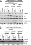
DDK interaction mutants of Claspin can still mediate activation of Chk1 and associate with replicating chromatin. A, nuclei were isolated from interphase extracts supplemented with 3,000 sperm nuclei/μl that were either immunodepleted with anti-Claspin antibodies (lanes 4–6 and 8–10) or mock depleted with control antibodies (lanes 7 and 11). The extracts were also incubated in the absence (lanes 4–7) or presence of aphidicolin (lanes 8–11). The extracts were supplemented with full-length versions of wild-type His6-Claspin-FLAG (lanes 5 and 9), His6-Claspin-FLAG-E/K (lanes 6 and 10), or no recombinant protein (lanes 4 and 8). Lanes 12–15 depict 4% of the extracts taken before isolation of nuclei. Aliquots of extracts were taken before (lane 1) and after immunodepletion (lanes 2 and 3). The extracts and nuclear fractions were immunoblotted for Claspin, Drf1, Mcm2, phosphorylated Ser344 of Chk1, and Chk1. B, chromatin was isolated from interphase extracts supplemented with 3,000 sperm nuclei/μl and incubated in the absence (lanes 4–8) or presence of aphidicolin (lanes 9–13). The extracts were immunodepleted with either anti-Claspin (lanes 4–7 and 9–12) or control antibodies (lanes 8 and 13). The extracts were also supplemented with the indicated versions of the His6-Claspin-FLAG protein (lanes 5–7 and 10–12) or buffer alone (lanes 4, 8, 9, and 13). Lanes 14–18 depict 4% of the extract taken before isolation of chromatin. Aliquots of extract were taken before (lane 1) and after immunodepletion (lanes 2 and 3). The samples were immunoblotted for Claspin, Cdc45, and Orc2.
FIGURE 6.
FIGURE 6.
Egg extracts containing DDK interaction mutants of Claspin replicate chromosomal DNA more slowly. Claspin-depleted extracts were prepared and supplemented with recombinant wild-type (WT, closed squares), D861E (open triangles), or E/K (asterisks) His6-Claspin-FLAG proteins or buffer alone (open circles). Recombinant Claspin proteins were added back at approximately the concentration of endogenous Claspin in egg extracts (see supplemental Fig. S6). Mock depleted extracts (closed diamonds) were prepared in parallel. Aliquots of extracts were taken at 35, 55, 75, and 95 min and assayed for chromosomal DNA replication. Incorporation of 32P into chromosomal DNA was determined by agarose gel electrophoresis and phosphorimaging. Incorporation was normalized to the level observed in the mock depleted extracts at 95 min. The data points represent the means ± S.E. for seven independent experiments.

Similar articles

Cited by

References

    1. Takeda D. Y., Dutta A. (2005) Oncogene 24, 2827–2843 - PubMed
    1. Arias E. E., Walter J. C. (2007) Genes Dev. 21, 497–518 - PubMed
    1. Kumagai H., Sato N., Yamada M., Mahony D., Seghezzi W., Lees E., Arai K., Masai H. (1999) Mol. Cell. Biol. 19, 5083–5095 - PMC - PubMed
    1. Jiang W., McDonald D., Hope T. J., Hunter T. (1999) EMBO J. 18, 5703–5713 - PMC - PubMed
    1. Montagnoli A., Bosotti R., Villa F., Rialland M., Brotherton D., Mercurio C., Berthelsen J., Santocanale C. (2002) EMBO J. 21, 3171–3181 - PMC - PubMed

Publication types

MeSH terms

LinkOut - more resources