. 2010 Apr 23;285(17):12803-12.
doi: 10.1074/jbc.M110.106989. Epub 2010 Feb 27.

A proteome-wide analysis of kinase-substrate network in the DNA damage response

Affiliations

A proteome-wide analysis of kinase-substrate network in the DNA damage response

Sheng-hong Chen et al. J Biol Chem. .

Abstract

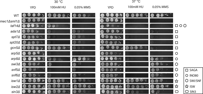
The DNA damage checkpoint, consisting of an evolutionarily conserved protein kinase cascade, controls the DNA damage response in eukaryotes. Knowledge of the in vivo substrates of the checkpoint kinases is essential toward understanding their functions. Here we used quantitative mass spectrometry to identify 53 new and 34 previously known targets of Mec1/Tel1, Rad53, and Dun1 in Saccharomyces cerevisiae. Analysis of replication protein A (RPA)-associated proteins reveals extensive physical interactions between RPA-associated proteins and Mec1/Tel1-specific substrates. Among them, multiple subunits of the chromatin remodeling complexes including ISW1, ISW2, INO80, SWR1, RSC, and SWI/SNF are identified and they undergo DNA damage-induced phosphorylation by Mec1 and Tel1. Taken together, this study greatly expands the existing knowledge of the targets of DNA damage checkpoint kinases and provides insights into the role of RPA-associated chromatins in mediating Mec1 and Tel1 substrate phosphorylation in vivo.


Figures

FIGURE 1.
FIGURE 1.
Quantitative MS identifies the targets of DNA damage checkpoint kinases. A, strategy used in identification of kinase-specific phosphopeptides (see “Experimental Procedures” for details). B, abundance ratio as a function of the integrated intensity for each phosphopeptide identified in the Mec1/Tel1 screen. A log10 scale is used for the integrated ion intensity of each heavy Lys/Arg-labeled phosphopeptide, whereas a log4 scale is used for the corresponding abundance ratio of heavy over light Lys/Arg-labeled phosphopeptide. C, steps toward identification of kinase-specific targets. A 4-fold cutoff was used to determine kinase-dependent phosphopeptides from the Mec1/Tel1, Rad53, and Dun1 screens, whose overlaps are analyzed using Venn diagrams (see text for further details).
FIGURE 2.
FIGURE 2.
Summary of the results from large-scale MS screens. A, comparison between the present study, a previous study by Smolka et al. (10), and all other studies. B, functional classification of the kinase-specific targets identified in our present study (also see Table 1). C, a partial summary of Mec1/Tel1-specific targets are colored in red, Rad53-specific targets are colored in green, and Dun1-specific targets are colored in blue. Solid lines indicate the known physical interactions between these targets.
FIGURE 3.
FIGURE 3.
Quantitative analysis of RPA-specific associated proteins. A, strategy used to identify specific associated proteins of RPA using Rfa1-TAP cells and the SILAC method. B, silver staining of 5% of the one-step affinity purified RPA compared with the mock purified sample. Arrows indicate specific bands corresponding to Rfa1 and Rfa2. C, examples of MS data of a representative peptide derived from each RPA-specific binding protein. In each case, the corresponding light isotope containing peptide is below the detection limit, whereas the heavy Lys/Arg containing peptide is detected. Asterisks indicate the natural occurrences of C13/N15 in the peptide, as revealed by high resolution MS. D, summary of RPA-associated proteins (indicated by filled and open circles, also see supplemental Table S5), along with Mec1/Tel1 substrates identified form the large-scale MS screens (indicated by a star). Solid lines indicate known physical interactions between them. Filled circles indicate the proteins found as both RPA-specific and Mec1/Tel1 substrates. Open circles indicate those only found as RPA-specific associated proteins.
FIGURE 4.
FIGURE 4.
Characterization of MMS-induced SQ/TQ phosphorylation of RPA, TFIID-SAGA, ISW1, ISW2, SWI/SNF, RSC, SWR1, and INO80 complexes. Only SQ/TQ phosphorylation sites of each protein complex and its associated proteins are shown. The numbers indicate the position of the phosphorylated serine or threonine in each protein. Solid lines indicate known physical associations between them. Filled circles indicate the phosphorylation site being detected as both Mec1/Tel1-specific from the large-scale MS screens and MMS-induced from the protein complex analysis, whereas open circles indicate that this phosphorylation is only found to be MMS-induced from protein complex analysis. For protein complex purification, TAP-tagged yeast strains are used as follows: A, Rfa1-TAP; B, Taf9-TAP; C, Isw1-TAP; D, Isw2-TAP; E, Ies1-TAP; F, Swr1-TAP; G, Sth1-TAP; H, Swi2-TAP.
FIGURE 5.
FIGURE 5.
DNA damage sensitivities of a subset of null mutants of various subunits of chromatin remodeling complexes (also see supplemental Fig. S1). A serial dilution of 5-fold was used to spot cells on the plates, which were then incubated at the indicated temperatures.

Similar articles

Cited by

References

    1. Harrison J. C., Haber J. E. (2006) Annu. Rev. Genet 40, 209–235 - PubMed
    1. Kolodner R. D., Putnam C. D., Myung K. (2002) Science 297, 552–557 - PubMed
    1. Nyberg K. A., Michelson R. J., Putnam C. W., Weinert T. A. (2002) Annu. Rev. Genet 36, 617–656 - PubMed
    1. Olsen J. V., Blagoev B., Gnad F., Macek B., Kumar C., Mortensen P., Mann M. (2006) Cell 127, 635–648 - PubMed
    1. Ong S. E., Blagoev B., Kratchmarova I., Kristensen D. B., Steen H., Pandey A., Mann M. (2002) Mol. Cell Proteomics 1, 376–386 - PubMed

Publication types

MeSH terms