

Localization and imaging of sialylated glycosphingolipids in brain tissue sections by MALDI mass spectrometry
- PMID: 20190299
- PMCID: PMC2900884
- DOI: 10.1093/glycob/cwq031
Localization and imaging of sialylated glycosphingolipids in brain tissue sections by MALDI mass spectrometry
Abstract
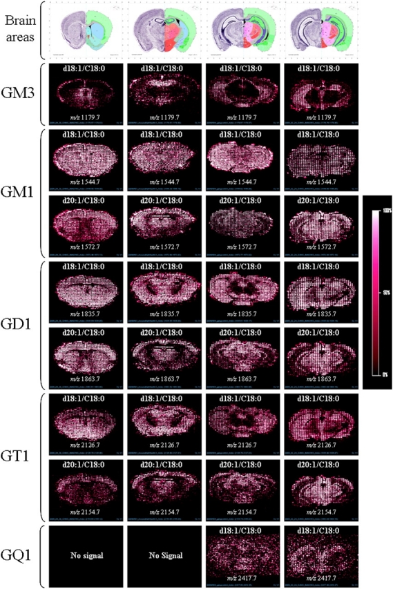
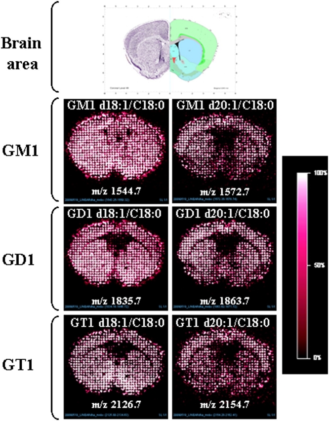
In this study, we describe a simple and efficient method for mapping the distribution and localization of all sialylated sphingoglycolipids present in coronal mouse brain sections using a conventional axial matrix-assisted laser desorption/ionization time of flight. A single scan of a histological tissue section gives a complete profile of ganglioside species without derivatization or labeling. We have developed and tested a new matrix preparation (2,6-dihydroxyacetophenone [DHA]/ammonium sulfate/heptafluorobutyric acid [HFBA]) to maximize the detection of all ganglioside species; the ammonium sulfate limits the formation of salt adducts, while the addition of HFBA increases the stability of DHA in a vacuum, thus facilitating imaging applications. Our results, in both extracted samples and whole tissue sections using negative ion reflectron and linear modes, show differences in localization in several brain regions depending on the sialic acids and the ceramide-associated core gangliosides.
Figures


References
-
- Caprioli RM, Farmer TB, Gile J. Molecular imaging of biological samples: localization of peptides and proteins using MALDI-TOF MS. Anal Chem. 1997;69(23):4751–4760. - PubMed
-
- Chan K, Lantier P, Liu X, Sandhu JK, Stanimirovic D, Li J. MALDI mass spectrometry imaging of gangliosides in mouse brain using ionic liquid matrix. Anal Chim Acta. 2009;639(1-2):57–61. - PubMed
-
- Chen Y, Allegood J, Liu Y, Wang E, Cachón-Gonzalez B, Cox TM, Merrill AH, Jr, Sullards MC. Imaging MALDI mass spectrometry using an oscillating capillary nebulizer matrix coating system and its application to analysis of lipids in brain from a mouse model of Tay-Sachs/Sandhoff disease. Anal Chem. 2008;80(8):2780–2788. - PubMed
-
- Cornett DS, Reyzer ML, Chaurand P, Caprioli RM. MALDI imaging mass spectrometry: molecular snapshots of biochemical systems. Nat Meth. 2007;4(10):828–833. - PubMed
