Review
. 2010 Mar;10(1):12-24.

Dynamic skeletal muscle stimulation and its potential in bone adaptation

Affiliations
Review

Dynamic skeletal muscle stimulation and its potential in bone adaptation

Y X Qin et al. J Musculoskelet Neuronal Interact. 2010 Mar.

Abstract

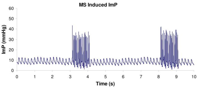
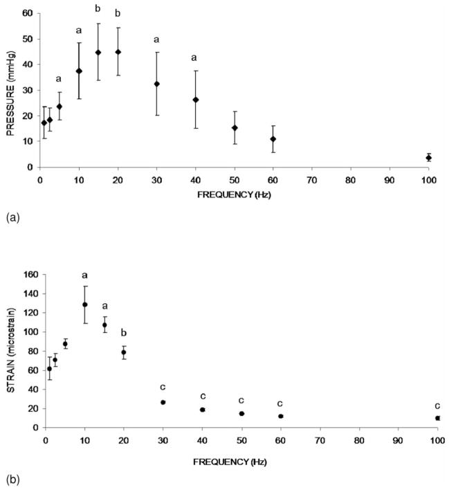
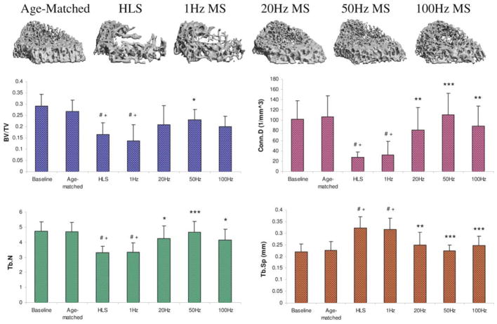
To identify mechanotransductive signals for combating musculoskeletal deterioration, it is essential to determine the components and mechanisms critical to the anabolic processes of musculoskeletal tissues. It is hypothesized that the interaction between bone and muscle may depend on fluid exchange in these tissues by mechanical loading. It has been shown that intramedullary pressure (ImP) and low-level bone strain induced by muscle stimulation (MS) has the potential to mitigate bone loss induced by disuse osteopenia. Optimized MS signals, i.e., low-intensity and high frequency, may be critical in maintaining bone mass and mitigating muscle atrophy. The objectives for this review are to discuss the potential for MS to induce ImP and strains on bone, to regulate bone adaptation, and to identify optimized stimulation frequency in the loading regimen. The potential for MS to regulate blood and fluid flow will also be discussed. The results suggest that oscillatory MS regulates fluid dynamics with minimal mechanical strain in bone. The response was shown to be dependent on loading frequency, serving as a critical mediator in mitigating bone loss. A specific regimen of dynamic MS may be optimized in vivo to attenuate disuse osteopenia and serve as a biomechanical intervention in the clinical setting.


Conflict of interest statement

The authors have no conflict of interest.

Figures

Figure 1
Figure 1
Intramedullary pressure generated by dynamic electrical muscle stimulation at 20Hz. Stimulation was applied for 1 seconds followed by 4 seconds rest. Normal heart beat generated approximately 4 mmHg of ImP (mean) in the femur. Muscle contraction increased ImP to 45 mmHg in a rat femoral model. (Courtesy of J Biomechanics 2009;42:140–5).
Figure 2
Figure 2
(a). Graphs show mean SD values from the ImP measurement. ImP in femur increased significantly with electrical frequency at 5, 10, 15, 20, 30, and 40 Hz. In the loading spectrum from 1 to 100 Hz, stimulation at 1 Hz generated an ImP of 18 mmHg. A maximum ImP of 45 mmHg was measured at 20 Hz, which was 2.5 folds higher than 1 Hz. ap<0.05 vs. baseline ImP; bp<0.01 vs. baseline ImP. (Courtesy of J Biomechanics 2009;42:140–5). (b). Graphs show mean SD values from the bone surface strain measurement. Dynamic muscle stimulation applied at various frequencies significantly increased bone strain. In the loading spectrum from 1 to 100 Hz, stimulation at 1 Hz produced a strain of 62 με. Peak strain of 128 was recorded at 10 Hz stimulation. The strain magnitude was reduced by >75% of the peak strain for stimulation frequencies greater than 30 Hz. ap<0.01 vs. 1, 2.5, and 5 Hz; bp<0.01 vs. 10 Hz; cp<0.001 vs. stimulation 20 Hz and below. (Courtesy of J Biomechanics 2009;42:140–5).
Figure 3
Figure 3
Trabecular bone at three metaphyseal sections and one epiphyseal region of the distal femurs was evaluated using microcomputed tomography. GP=growth plate (arrow); M=metaphysis; E=epiphysis. (Courtesy of BONE 2008;43:1093–1100).
Figure 4
Figure 4
Representative 3D μCT images of trabecular bone in the M3 region (750 μm, immediately above the growth plate). Graphs show mean + SD values for bone volume fraction (BV/TV, %), connectivity density (Conn.D, 1/mm3), trabecular number (Tb.N, 1/mm), and separation (Tb.Sp, mm) at the M3 region. Only 50 Hz MS demonstrated significant preventive effects for all indices against 4-week HLS. #p<0.001 vs. baseline; +p<0.001 vs. age-matched; *p<0.05 vs. HLS & 1 Hz MS; **p<0.01 vs. HLS & 1 Hz MS; ***p<0.001 vs. HLS & 1 Hz MS. (Courtesy of BONE 2008;43:1093–100).
Figure 5
Figure 5
Graphs show the percentage differences between HLS and MS experimental groups SD values in all three metaphyseal regions for bone volume fraction (BV/TV) and connectivity density (Conn.D). For MS with mid to high stimulation frequencies, the levels of effectiveness on trabecular bone against functional disuse alone were always greatest at M1 and least at M3. **p<0.001 vs. M3; *p<0.05 vs. M3; #p<0.001 vs. M2. (Courtesy of BONE 2008;43:1093–1100).

Similar articles

Cited by

References

    1. Akima H, Katayama K, Sato K, Ishida K, Masuda K, Takada H, Watanabe Y, Iwase S. Intensive cycle training with artificial gravity maintains muscle size during bed rest. Aviat Space Environ Med. 2005;76:923–9. - PubMed
    1. Akima H, Kawakami Y, Kubo K, Sekiguchi C, Ohshima H, Miyamoto A, Fukunaga T. Effect of short-duration spaceflight on thigh and leg muscle volume. Med Sci Sports Exerc. 2000;32:1743–7. - PubMed
    1. Allen MR, Hogan HA, Bloomfield SA. Differential bone and muscle recovery following hindlimb unloading in skeletally mature male rats. J Musculoskelet Neuronal Interact. 2006;6:217–25. - PubMed
    1. BeDell KK, Scremin AM, Perell KL, Kunkel CF. Effects of functional electrical stimulation-induced lower extremity cycling on bone density of spinal cord-injured patients. Am J Phys Med Rehabil. 1996;75:29–34. - PubMed
    1. Belanger M, Stein RB, Wheeler GD, Gordon T, Leduc B. Electrical stimulation: can it increase muscle strength and reverse osteopenia in spinal cord injured individuals? Arch Phys Med Rehabil. 2000;81:1090–8. - PubMed

Publication types

MeSH terms