Review
. 2010 Mar;10(1):35-45.

The development and morphogenesis of the tendon-to-bone insertion - what development can teach us about healing -

Affiliations
Review

The development and morphogenesis of the tendon-to-bone insertion - what development can teach us about healing -

S Thomopoulos et al. J Musculoskelet Neuronal Interact. 2010 Mar.

Abstract

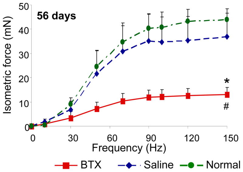
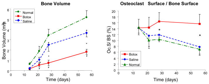
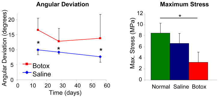
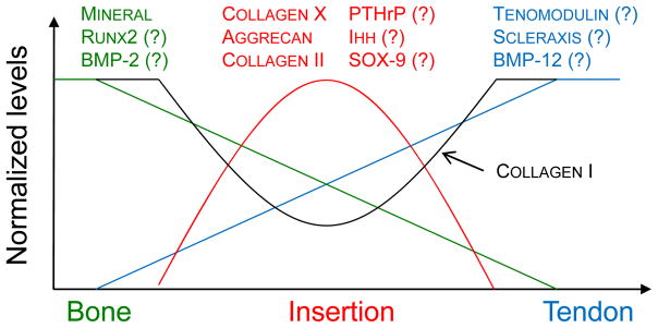
The attachment of dissimilar materials is a major challenge because of the high levels of stress that develop at such interfaces. An effective solution to this problem develops at the attachment of tendon (a compliant "soft tissue") to bone (a stiff "hard tissue"). This tissue, the "enthesis", transitions from tendon to bone through gradations in structure, composition, and mechanical properties. These gradations are not regenerated during tendon-to-bone healing, leading to a high incidence of failure after surgical repair. Understanding the development of the enthesis may allow scientists to develop treatments that regenerate the natural tendon-to-bone insertion. Recent work has demonstrated that both biologic and mechanical factors drive the development and morphogenesis of the enthesis. A cascade of biologic signals similar to those seen in the growth plate promotes mineralization of cartilage on the bony end of the enthesis and the formation of fibrocartilage on the tendon end of the enthesis. Mechanical loading is also necessary for the development of the enthesis. Removal of muscle load impairs the formation of bone, fibrocartilage, and tendon at the developing enthesis. This paper reviews recent work on the development of the enthesis, with an emphasis on the roles of biologic and mechanical factors.


Figures

Figure 1
Figure 1
Morphology of the supraspinatus tendon-to-bone insertion site.
Figure 2
Figure 2
There is a gradual change in the degree of mineralization across the tendon-to-bone insertion of the rotator cuff as evaluated by Raman spectroscopy (right panel, apatite [960 Δcm−1] to collagen [2940 Δcm−1] peak ratios are plotted relative to position. [Adapted with permission from Wopenka B, Kent A, Pasteris JD, Yoon Y, Thomopoulos S. The Tendon-to-Bone Transition of the Rotator Cuff: A Preliminary Raman Spectroscopic Study Documenting the Gradual Mineralization Across the Insertion in Rat Tissue Samples. Appl Spectrosc. 2008 Dec;62(12):1285–94.]
Figure 3
Figure 3
Bounds (lines) and Monte Carlo estimates (circles) for the elastic modulus (E) of collagen fibrils containing mineral deposits up to the level of mineralization found in bone. The stiffening of collagen fibrils by mineral increases dramatically above a critical mineral concentration called the “percolation threshold” (arrows). This concentration is a function of the shape and distribution of mineral (red: aspect ratio of 1:1; blue: aspect ratio of 2:1). [Adapted with permission from Genin GM, Kent A, Birman V, Wopenka B, Pasteris JD, JP M, Thomopoulos S. Functional grading of mineral and collagen in the attachment of tendon to bone. Biophysical Journal. 2009;97(4).]
Figure 4
Figure 4
Serial sections for collagen I (A–E) and collagen II (F–J) expression at 13.5 dpc (A, F), 18.5 dpc (B, G), neonatal (C, H), 7 days (D, I), and 28 days (E, J) (Toluidine blue stain). Type I collagen expression mirrored type II collagen expression across the insertion site. Type I collagen was always found on the tendinous side of the insertion, and type II collagen was always found on the bony side of the insertion. Positive staining is indicated by black grains. s, supraspinatus tendon; h, humeral head; i, interface. [Adapted with permission from Galatz L, Rothermich S, VanderPloeg K, Petersen B, Sandell L, Thomopoulos S. Development of the supraspinatus tendon-to-bone insertion: localized expression of extracellular matrix and growth factor genes. J Orthop Res. 2007 Dec;25(12):1621–8.]
Figure 5
Figure 5
Expression of Patched-1, the receptor for Ihh, was localized to the developing enthesis (21 days postnatally, t: tendon, b: bone, i: insertion, scale bar = 100μm).
Figure 6
Figure 6
The contractile properties of BTX muscles were significantly lower compared to the Saline and Normal muscles (* p <0.05, paired t-test, BTX vs. Saline; # p <0.05, BTX or Saline vs. Normal).
Figure 7
Figure 7
Bone volume was similar when comparing Botox to Saline at 14 days. Bone volume, however, was significantly different when comparing Botox to Saline at 21, 28, and 56 days. The percentage of bone surface covered with osteoclasts (Oc.S/BS) was similar in all groups at 14 days but significantly different when comparing Saline or Normal to the Botox group at 21–56 days. [* Significantly different- Botox vs. Saline, # Significantly different- Botox/Saline vs. Normal, p < 0.05]. [Adapted with permission from Thomopoulos S, Kim HM, Rothermich SY, Biederstadt C, Das R, Galatz LM. Decreased muscle loading delays maturation of the tendon enthesis during postnatal development. J Orthop Res. 2007 Sep;25(9):1154–63.]
Figure 8
Figure 8
Tendon angular deviation was significantly higher (i.e., the collagen fiber distribution was less organized) in the Botox group compared to the Saline group. Ultimate stress was significantly lower in the Botox group compared to the Saline and Normal groups (* p < 0.05).
Figure 9
Figure 9
Gradations in biologic factors may promote gradations in cell differentiation and subsequent tissue formation. The measured gradations in mineral at the mature enthesis (shown in green, highest in bone and lowest in tendon) may be due to a graded expression of osteogenic factors such Runx2 and BMP-2. The measured gradations in fibrocartilage (shown in red, low in both tendon and bone and highest at the insertion) may be due to a graded expression of factors such as PTHrP, Ihh, and Sox9. Differences in tenogenesis (shown in blue, highest in tendon and lowest in bone) may be due a graded expression of tendon specific factors such as scleraxis, BMP-12, and tenomodulin.

Similar articles

Cited by

References

    1. Bostrom MPG, Boskey A, Kauffman JK, Einhorn TA. Form and function of bone. In: Buckwalter JA, Einhorn TA, Simon SR, editors. Orthopaedic Basic Science. 2. AAOS; 2000. pp. 319–70.
    1. Woo SL, An K, Frank CB, Livesay GA, Ma CB, Zeminski JA, Wayne JS, Myers BS. Orthopaedic Basic Science. 2. Rosemont, IL: AAOS; 2000. Anatomy, biology, and biomechanics of tendon and ligament; pp. 581–616.
    1. Jones RM. Mechanics of composite materials. 2. Philadelphia: Taylor & Francis, Inc; 1999.
    1. Williams ML. Stress singularities resulting from various boundary conditions in angular corners of plates in extension. Journal of Applied Mechanics. 1952;19:526–8.
    1. Aoki M, Oguma H, Fukushima S, Ishii S, Ohtani S, Murakami G. Fibrous connection to bone after immediate repair of the canine infraspinatus: the most effective bony surface for tendon attachment.[see comment] Journal of Shoulder & Elbow Surgery. 2001;10(2):123–8. - PubMed

Publication types

MeSH terms