. 2010 Apr;12(4):372-9.
doi: 10.1038/ncb2037. Epub 2010 Feb 28.

Genome-wide RNA-mediated interference screen identifies miR-19 targets in Notch-induced T-cell acute lymphoblastic leukaemia

Affiliations

Genome-wide RNA-mediated interference screen identifies miR-19 targets in Notch-induced T-cell acute lymphoblastic leukaemia

Konstantinos J Mavrakis et al. Nat Cell Biol. 2010 Apr.

Abstract

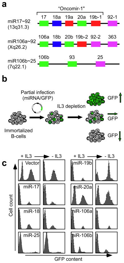
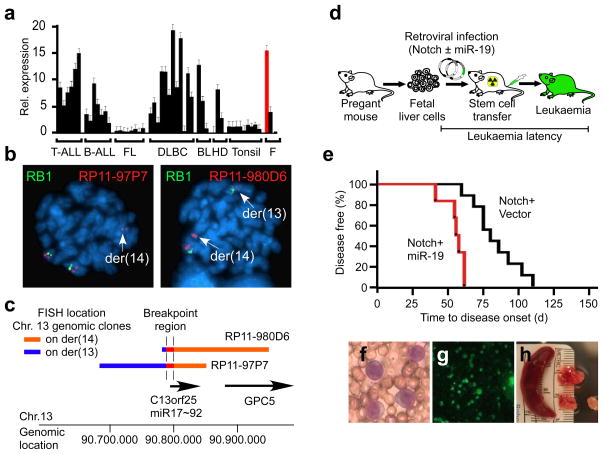
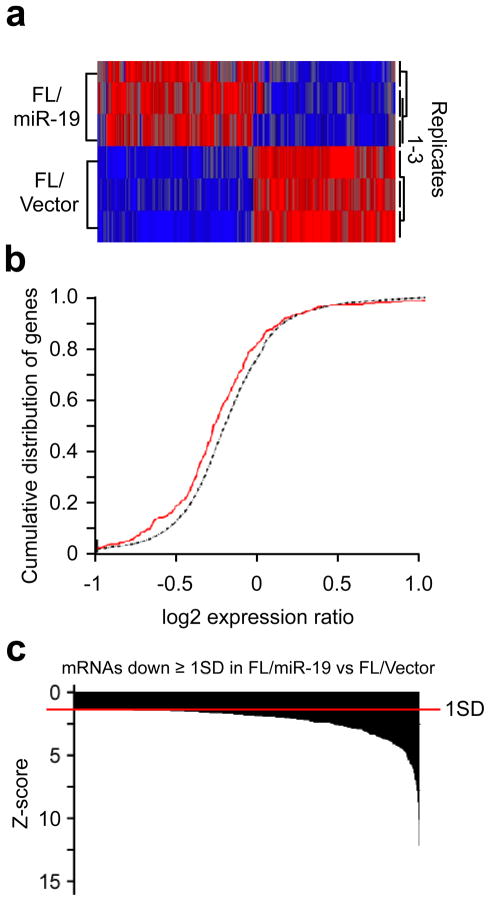
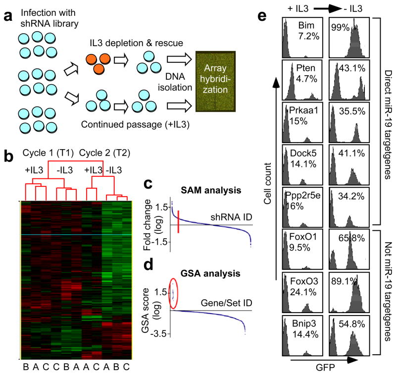
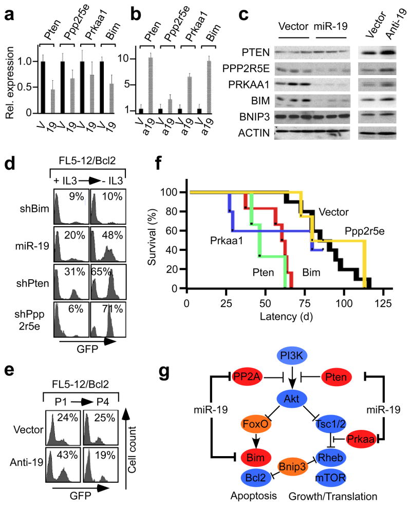
MicroRNAs (miRNAs) have emerged as novel cancer genes. In particular, the miR-17-92 cluster, containing six individual miRNAs, is highly expressed in haematopoietic cancers and promotes lymphomagenesis in vivo. Clinical use of these findings hinges on isolating the oncogenic activity within the 17-92 cluster and defining its relevant target genes. Here we show that miR-19 is sufficient to promote leukaemogenesis in Notch1-induced T-cell acute lymphoblastic leukaemia (T-ALL) in vivo. In concord with the pathogenic importance of this interaction in T-ALL, we report a novel translocation that targets the 17-92 cluster and coincides with a second rearrangement that activates Notch1. To identify the miR-19 targets responsible for its oncogenic action, we conducted a large-scale short hairpin RNA screen for genes whose knockdown can phenocopy miR-19. Strikingly, the results of this screen were enriched for miR-19 target genes, and include Bim (Bcl2L11), AMP-activated kinase (Prkaa1) and the phosphatases Pten and PP2A (Ppp2r5e). Hence, an unbiased, functional genomics approach reveals a coordinate clampdown on several regulators of phosphatidylinositol-3-OH kinase-related survival signals by the leukaemogenic miR-19.


Conflict of interest statement

Author information The authors declare no competing financial interests.

Figures

Figure 1
Figure 1. miR-19 enhances cytokine independent survival in vitro
a, Genomic organization of the 17~92 cluster (including “oncomir-1”) and its paralogues, miRNAs shown in identical colour share common seed sequences; b, Schematic of the competition assay for cytokine independent survival of immortalized FL5-12 lymphocytes; c, Representative FACS profiles showing enrichment of miR-19/GFP expressing FL5-12 cells upon IL3 depletion, while populations transduced with vector or the other miRNAs remain unchanged (all experiments in triplicates).
Figure 2
Figure 2. miR-19 is a novel T-ALL oncogene
a, qRT-PCR measurement of miR-19 expression in a panel of human lymphoid malignancies: (T-/B-ALL) T- and B-cell acute lymphatic leukaemia; (FL) follicular lymphoma; (DLBCL) diffuse large B-cell lymphoma; (BL) Burkitt’s lymphoma; (HD) Hodgkin’s disease; (Tonsil) lymphocytes from reactive tonsils; (F) indicates FL5-12 cells, both parental (black bar) and miR-19 transduced (red bar) shown are mean +/− SD; b, Double colour FISH analysis of t(13;14)(q32;q11) using a RB1 probe (green) in 13q14 and genomic clones RP11-97P7 and RP11-980D6 overlapping the 17~92 locus in 13q32 (red). c, Graphic representation of FISH results; d, Mouse model of Notch-induced T-ALL; e, Kaplan-Meier analysis of leukaemia free survival after HPC transplantation (Red: Notch-ICN + miR-19; n=6; black: Notch-ICN + vector; n=9); f-h, Representative microphotographs of Notch/miR-19 induced ALL; f, Leukaemic blasts on blood film; g) Effacement of the bone marrow by miR-19/GFP expressing leukaemic cells; h) Splenomegaly and lymphomas. The pathologic appearance of Notch1 induced leukaemia is identical (not shown).
Figure 3
Figure 3. Gene expression analysis of parental and miR-19 transduced FL5-12 cells
a, Heat-map illustration of the unsupervised clustering analysis reveals differences in gene expression between parental (FL/Vector) and miR-19 expressing FL5-12 cells (FL/miR-19); b, Comparison of the expression change of predicted miR-19 targets represented on the array (336 genes, red line) versus all represented genes (8065 genes, black line) (p < 2e-04; KS-test); c, Histogram of genes whose expression is down regulated by > 1SD in FL5-12/miR-19 cells compared to parental cells: miR-19 target genes are not over represented in the among genes showing more pronounced (1.5 or 2 SD) reductions in expression. Expression array readily detects global down regulation by miR-19, but is not a sufficient filter to define key miR -19 targets.
Figure 4
Figure 4. Genetic screen for shRNAs that phenocopy miR-19 in lymphocyte survival
a, Design schematic of the pooled shRNA screen, where large populations of FL5-12 cells are transduced with library pools and subjected to IL-3 depletion; b, Heat-map of the unsupervised clustering analysis of the half-hairpin array results. (T1, T2) indicate time points after 1 and 2 cycles of IL3 depletion; (-IL3) treated and (+IL3) untreated groups; (A-C) are the replicates (replicate B at T2/+IL3 was removed for technical reasons); c, Statistical analysis of Microarray data (SAM) indicating fold change (log2) for individual shRNAs and the threshold for validation studies (red line); d, Gene Set enrichment analysis (GSA) identifies enrichment of sets of shRNAs targeting 14 genes (red circle); e, Representative FACS profiles from triplicate validation experiments showing the enrichment of FL5-12 cells expressing the indicated shRNAs and GFP upon IL3 depletion. Vector alone showed no change (not shown).
Figure 5
Figure 5. The identified genes are actual and relevant targets of miR-19
a, qRT-PCR for the indicated genes on cDNA prepared from vector (V; black bars) or miR-19 transduced (19; shaded bars) FL5-12 cells. Expression levels (mean +/− SD) are normalized to the vector controls (relative expression); b, qRT-PCR comparing FL5-12 cells transduced with vector (V; black bars) and antagomirs to miR-19 (a19; shaded bars) analyzed as above; c, Immunoblot on lysates from vector, miR-19 or antagomir (Anti-19) transduced FL5-12 cells probed for the indicated proteins (see also Suppl. Fig. 17); d, Representative FACS profiles of triplicate experiments on FL5-12 cells co-expressing Bcl2 (FL5-12/Bcl2) and the indicated shRNAs or miR-19 and GFP. Cells are shifted from complete (+IL3) to IL3 deficient media (-IL3) to assess Bcl2 independent effects on lymphocyte survival; e, FACS analysis of FL5-12/Bcl2 cells transduced with antagomir (Anti-19) or vector and GFP and grown in complete media; f, In vivo knockdown of individual target genes accelerates Notch1-induced T-ALL in vivo. Kaplan-Meier analysis of survival after HPC transplantation (Black: vector n=10; green: shPten n=4; red: shBim n=6; blue: shPrkaa n=5; yellow: shPp2r5e n=4); g, Diagram indicating the multilevel control of PI3K survival signals by miR-19.

Similar articles

Cited by

References

    1. Xiao C, et al. Lymphoproliferative disease and autoimmunity in mice with increased miR-17-92 expression in lymphocytes. Nat Immunol. 2008;9:405–414. - PMC - PubMed
    1. He L, et al. A microRNA polycistron as a potential human oncogene. Nature. 2005;435:828–833. - PMC - PubMed
    1. Ventura A, et al. Targeted deletion reveals essential and overlapping functions of the miR-17 through 92 family of miRNA clusters. Cell. 2008;132:875–886. - PMC - PubMed
    1. Bartel DP. MicroRNAs: genomics, biogenesis, mechanism, and function. Cell. 2004;116:281–297. - PubMed
    1. Friedman RC, Farh KK, Burge CB, Bartel DP. Most mammalian mRNAs are conserved targets of microRNAs. Genome Res. 2009;19:92–105. - PMC - PubMed

Publication types

MeSH terms