Nanostructured films from hierarchical self-assembly of amyloidogenic proteins
- PMID: 20190750
- PMCID: PMC4612398
- DOI: 10.1038/nnano.2010.26
Nanostructured films from hierarchical self-assembly of amyloidogenic proteins
Abstract
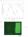
In nature, sophisticated functional materials are created through hierarchical self-assembly of simple nanoscale motifs. In the laboratory, much progress has been made in the controlled assembly of molecules into one-, two- and three-dimensional artificial nanostructures, but bridging from the nanoscale to the macroscale to create useful macroscopic materials remains a challenge. Here we show a scalable self-assembly approach to making free-standing films from amyloid protein fibrils. The films were well ordered and highly rigid, with a Young's modulus of up to 5-7 GPa, which is comparable to the highest values for proteinaceous materials found in nature. We show that the self-organizing protein scaffolds can align otherwise unstructured components (such as fluorophores) within the macroscopic films. Multiscale self-assembly that relies on highly specific biomolecular interactions is an attractive path for realizing new multifunctional materials built from the bottom up.
Figures


Comment in
-
Strength in numbers.Nat Nanotechnol. 2010 Mar;5(3):172-4. doi: 10.1038/nnano.2010.28. Epub 2010 Feb 28. Nat Nanotechnol. 2010. PMID: 20190749 No abstract available.
Similar articles
-
Characterization of the nanoscale properties of individual amyloid fibrils.Proc Natl Acad Sci U S A. 2006 Oct 24;103(43):15806-11. doi: 10.1073/pnas.0604035103. Epub 2006 Oct 12. Proc Natl Acad Sci U S A. 2006. PMID: 17038504 Free PMC article.
-
Self-folding and aggregation of amyloid nanofibrils.Nanoscale. 2011 Apr;3(4):1748-55. doi: 10.1039/c0nr00840k. Epub 2011 Feb 23. Nanoscale. 2011. PMID: 21347488
-
Imaging Protein Fibers at the Nanoscale and In Situ.Methods Mol Biol. 2018;1777:83-100. doi: 10.1007/978-1-4939-7811-3_4. Methods Mol Biol. 2018. PMID: 29744829
-
Nanomechanical strength mechanisms of hierarchical biological materials and tissues.Comput Methods Biomech Biomed Engin. 2008 Dec;11(6):595-607. doi: 10.1080/10255840802078030. Comput Methods Biomech Biomed Engin. 2008. PMID: 18803059 Review.
-
Spatially-interactive biomolecular networks organized by nucleic acid nanostructures.Acc Chem Res. 2012 Aug 21;45(8):1215-26. doi: 10.1021/ar200295q. Epub 2012 May 29. Acc Chem Res. 2012. PMID: 22642503 Free PMC article. Review.
Cited by
-
Protein nanofibrils and their use as building blocks of sustainable materials.RSC Adv. 2021 Dec 8;11(62):39188-39215. doi: 10.1039/d1ra06878d. eCollection 2021 Dec 6. RSC Adv. 2021. PMID: 35492452 Free PMC article. Review.
-
The emerging field of RNA nanotechnology.Nat Nanotechnol. 2010 Dec;5(12):833-42. doi: 10.1038/nnano.2010.231. Epub 2010 Nov 21. Nat Nanotechnol. 2010. PMID: 21102465 Free PMC article. Review.
-
From Fundamental Amyloid Protein Self-Assembly to Development of Bioplastics.Biomacromolecules. 2024 Jan 8;25(1):5-23. doi: 10.1021/acs.biomac.3c01129. Epub 2023 Dec 26. Biomacromolecules. 2024. PMID: 38147506 Free PMC article. Review.
-
Hybrid Spider Silk with Inorganic Nanomaterials.Nanomaterials (Basel). 2020 Sep 16;10(9):1853. doi: 10.3390/nano10091853. Nanomaterials (Basel). 2020. PMID: 32947954 Free PMC article. Review.
-
The Interplay between Whey Protein Fibrils with Carbon Nanotubes or Carbon Nano-Onions.Materials (Basel). 2021 Jan 28;14(3):608. doi: 10.3390/ma14030608. Materials (Basel). 2021. PMID: 33525699 Free PMC article.
References
-
- Dobson CM. Protein folding and misfolding. Nature. 2003;426:884–890. - PubMed
-
- Buehler MJ, Ackbarow T. Nanomechanical strength mechanisms of hierarchical biological materials and tissues. Comput. Methods Biomech. Biomed. Eng. 2008;11:595–607. - PubMed
-
- Buehler MJ, Yung YC. Deformation and failure of protein materials in physiologically extreme conditions and disease. Nature Mater. 2009;8:175–188. - PubMed
-
- Whitesides GM, Grzybowski B. Self-assembly at all scales. Science. 2002;295:2418–2421. - PubMed
-
- Kol N, et al. Self-assembled peptide nanotubes are uniquely rigid bioinspired supramolecular structures. Nano Lett. 2005;5:1343–1346. - PubMed
Publication types
MeSH terms
Substances
Grants and funding
LinkOut - more resources
Full Text Sources
Other Literature Sources

