. 2010 Mar;16(3):279-85.
doi: 10.1038/nm.2092. Epub 2010 Feb 28.

Kynurenine is an endothelium-derived relaxing factor produced during inflammation

Affiliations

Kynurenine is an endothelium-derived relaxing factor produced during inflammation

Yutang Wang et al. Nat Med. 2010 Mar.

Erratum in

  • Nat Med. 2010 May;16(5):607

Abstract

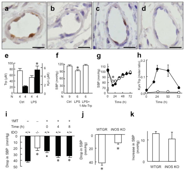
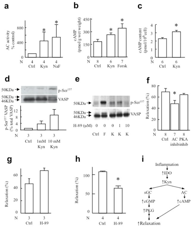
Control of blood vessel tone is central to vascular homeostasis. Here we show that metabolism of tryptophan to kynurenine by indoleamine 2,3-dioxygenase (Ido) expressed in endothelial cells contributes to arterial vessel relaxation and the control of blood pressure. Infection of mice with malarial parasites (Plasmodium berghei) or induction of endotoxemia in mice led to endothelial expression of Ido, decreased plasma tryptophan concentration, increased kynurenine concentration and hypotension. Pharmacological inhibition of Ido increased blood pressure in systemically inflamed mice but not in mice deficient in Ido or interferon-gamma, which is required for Ido induction. Both tryptophan and kynurenine dilated preconstricted porcine coronary arteries; the dilating effect of tryptophan required the presence of active Ido and an intact endothelium, whereas the effect of kynurenine was endothelium independent. The arterial relaxation induced by kynurenine was mediated by activation of the adenylate and soluble guanylate cyclase pathways. Kynurenine administration decreased blood pressure in a dose-dependent manner in spontaneously hypertensive rats. Our results identify tryptophan metabolism by Ido as a new pathway contributing to the regulation of vascular tone.


Figures

Fig. 1
Fig. 1. IDO pathway contributes to the regulation of BP in PbA-infected mice
(a-c) Expression of IDO in endothelial cells of resistance arteries in kidney. IDO expression was assessed by immunohistochemistry in non-infected mice and in mice 5 days post-infection with PbA (bar = 40 μm). IDO was expressed strongly in endothelial cells of PbA-infected mice (a) but was absent in non-infected mice (b) and in corresponding isotype control sections of PbA-infected mice (c). (d) Decreased plasma Trp and (e) increased Kyn concentration in PbA-infected Ido+/+ not Ido-/- mice. (f-g) SBP determined by tail cuff method in conscious C57BL/6J wild type (f) or Ido-/- (g) mice with or without PbA infection, before and after i.p. injection of 1 mL 20 mM 1-Me-Trp. Results in d-g represent mean±SEM, with the number of animals (N) used for each treatment indicated. *P<0.05 versus uninfected control (d, f) or before 1-Me-Trp injection in PbA-infected mice (g).
Fig. 2
Fig. 2. IDO contributes to the regulation of BP in LPS-induced endotoxemia
Expression of IDO in endothelial cells of resistance arteries of kidney (a,b) and intestine (c,d) assessed by immunohistochemistry in C57BL/6J mice 48 h after treatment with LPS (7.5 mg/kg, i.p.) (bar = 10 μm). IDO was expressed in endothelial cells of endotoxemic mice (a,c) but staining was absent in isotype controls (b,d). (e) Plasma concentration of Trp (open columns) and Kyn (filled columns) in endotoxemic mice 48 h after i.p. injection of LPS. *P<0.05 versus control. (f) SBP in endotoxemic mice 48 h after LPS injection. *P<0.05 versus both control and LPS+1-Me-Trp. (g) SBP in wild type (o) and Ido-/- mice (●) after LPS injection (n=12-30 for each time point). *P<0.01, wild type versus Ido-/- mice. (h) Ratio of plasma Kyn/Trp, a marker of IDO activity, in wild type and Ido-/- mice (n=3-5 for each time point) after LPS injection. (i) Drop in SBP caused by LPS in wild type versus Ido-/- mice, and in wild type mice co-administered with 1-Me-Trp (50 mg/kg, i.p.) at 0, 4, 8 h post LPS. (j) Drop in SBP of wild type (WTGR) and iNos-/- mice 24 h after LPS (n=4 animals per group). *P<0.05 versus untreated mice. (k) Increase in SBP by 1-Me-Trp in mice at 24 h after LPS injection (n=4 animals per group).
Fig. 3
Fig. 3. IFNγ-dependent expression of IDO in endothelial cells and its associated conversion of Trp to Kyn
(a-d) IFNγ induced expression of IDO protein and activity in porcine aortic endothelial cells. Cells were incubated for 20 h in the presence of Trp (200 μM) and the indicated concentration of IFNγ (a, b) or with 250 ng IFNγ/mL for the indicated times (c, d). Cells were harvested. Expression of IDO (46 KDa) and β-actin (42 KDa) was assessed by Western blot and quantified by densitometry (a, c). The concentration of Trp (circles) and Kyn (squares) in the supernate was determined (b, d) in the presence (filled symbols) and absence (open symbols) of cells. (e-f) Production of Kyn by porcine coronary arteries. Fresh porcine circumflex arteries were incubated at 37 °C for up to 24 h in M199 containing 200 μM Trp. (e) Arteries with intact endothelium (EC) were incubated with 0 (❍), 200 (▲) or 400 ng IFNγ/mL (■), the released Kyn determined and its concentration in the coronary lumen calculated taking into account the ratio of luminal versus medium volume. (f) Arteries with intact or denuded EC were incubated for 24 h in the absence or presence of 400 ng IFNγ/mL or the IDO inhibitor 1-Me-Trp (1 mM). Results show mean±SEM of N=3 (a-d) or 4 (e-f) independent experiments. Where error bars are not shown, symbol is larger than the error. *P<0.01 versus control (e) or all other groups (f); P<0.01 versus 200 ng IFNγ/mL (e).
Fig. 4
Fig. 4. The IDO pathway metabolite Kyn regulates vascular tone
(a) Relaxation of porcine coronary arteries in response to addition of Trp. Fresh porcine circumflex arteries with intact or denuded endothelium (EC) were incubated for 20 h at 37 °C in M199 in the absence or presence of 400 ng IFNγ/mL. Arteries were then removed, washed and incubated with 300 μM N-nitro-l-arginine methyl ester and 10 μM indomethacin for 30 min and pre-contracted with U-46619 to 50-60% of maximum force. Relaxation was determined in response to addition of Trp (8 mM final concentration). *P<0.05 versus EC-denuded and 1-Me-Trp-treated groups. (b) Contraction of porcine coronary artery in response to U-46619 in control preparations (❍) and in preparations pre-treated with 6 mM Kyn for 1 h (●). *P<0.05 versus control. (c) Relaxation of denuded vessel rings (▲) or rings with intact EC in response to added DMSO (❍, vehicle control) or Kyn (●). *P<0.05 versus control. (d, e) Trp, Kyn or sodium nitroprusside (SNP) was injected into spontaneously hypertensive rats via the femoral vein to give final blood concentrations ranging from 0.15 – 1.5 mM (for Kyn and Trp) and 0.15 – 1.5 μM (SNP), with systolic blood pressure being monitored continuously. We assumed a total blood volume of 17 mL for the 250 g rats used. (d) Typical blood pressure responses elicited by 1.5 mM of either Kyn or Trp, or 1.5 μM SNP. (e) Dose-dependent changes in systolic blood pressure following injection of Kyn (●), Trp (Δ) or SNP (❍). The results show mean±SEM of four (a), six (b, c) or three (e) independent experiments.
Fig. 5
Fig. 5. Kyn relaxes blood vessels via the sGC/cGMP/PKG pathway
(a) Activity of heme-containing (empty bars) and heme-free (filled bars) purified rat sGC in response to vehicle (control), 5 mM Kyn or 10 nM DEA/NO. (b) cGMP content in intact coronary arteries exposed to 1 mM Kyn, 10 μM ODQ or 1 mM Kyn + 10 μM ODQ in the presence of 300 μM nitro-l-arginine methyl ester and 10 μM indomethacin. (c) Response of porcine coronary arteries to 3 mM Kyn after pretreatment for 30 min with 10 μM ODQ or 10 μM Rp-8-CTP-cGMPS (inhibitor of cGMP-dependent protein kinase, PKG). (d) Activity of heme-containing sGC in response to Kyn in the absence (❍) or presence (●) of 10 μM ODQ. Pre-treatment of porcine coronary arteries with 10 μM ODQ (filled bars) at 37 °C for 24 h decreased (e) the content of sGCβ subunit, (f) vasorelaxation to DEA/NO and (g) Kyn, when compared with control (24 h incubation at 37 °C without ODQ, empty bars). (h) Enhancement of phosphorylation of vasodilator-stimulated phosphoprotein (VASP) at Ser239 in human platelets, after 3 min stimulation with 10 mM Kyn or 300 nM DEA/NO. Results show mean±SEM with N as indicated, except (d) and (e) that are representative of three separate experiments. Western blots (e) and (h) are representative of three separate experiments. *P≤0.05 versus respective control.
Fig. 6
Fig. 6. Involvement of the adenylyl cyclase/cAMP pathway in vessel relaxation induced by Kyn
(a) Activation of adenylyl cyclase by 1 mM Kyn or 10 mM NaF. Adenylyl cyclase was enriched as a membrane preparation from porcine coronary arteries and exposed to the agent indicated for 12 min at 37 °C before determination of cAMP. (b) cAMP levels in intact coronary arteries exposed to 1 mM Kyn or 0.6 μM forskolin (positive control) in the presence of isobutylmethylxanthine (inhibitor of phosphodiesterases). (c) cAMP levels in rat aortic smooth muscle cells exposed to vehicle (PBS, control) or 1 mM Kyn for 5 min. (d) Phosphorylation of VASP at Ser157 in human platelet after a 3-min stimulation with 1 or 10 mM Kyn. (e) VASP phosphorylation at Ser157 in MEG-01 cells 3 min after stimulation with 1 mM Kyn (K) in the presence or absence of the PKA inhibitor H-89 (0, 1 and 10 μM). Forskolin (F) was used as a positive control. (f) Response of porcine coronary arteries to 3 mM Kyn after pretreatment for 30 min with 100 μM SQ22,536 (inhibitor of adenylyl cyclase, AC) or 30 μM Rp-8-CTP-cAMPS (inhibitor of cAMP-dependent protein kinase, PKA). (g-h) Relaxation of pre-constricted mouse aortic rings to 3 mM Kyn (g) or 1 μM forskolin (h) after pretreatment for 30 min with 10 μM of the PKA inhibitor H-89. Results show mean±SEM with N as indicated. *P<0.05 versus respective control. (i) Proposed mechanism of inflammation-dependent contribution of Trp metabolism to vessel relaxation during systemic inflammation.

Comment in

Similar articles

Cited by

References

    1. Riedemann NC, Guo RF, Ward PA. Novel strategies for the treatment of sepsis. Nature Medicine. 2003;9:517–523. - PubMed
    1. Gomez-Jimenez J, et al. L-arginine: nitric oxide pathway in endotoxemia and human septic shock. Crit Care Med. 1995;23:253–258. - PubMed
    1. Ignarro LJ, Cirino G, Casini A, Napoli C. Nitric oxide as a signaling molecule in the vascular system: an overview. J Cardiovasc Pharmacol. 1999;34:879–886. - PubMed
    1. Kröncke KD, Fehsel K, Kolb-Bachofen V. Inducible nitric oxide synthase in human diseases. Clin Exp Immunol. 1998;113:147–156. - PMC - PubMed
    1. Meyer J, et al. Reversal of hyperdynamic response to continuous endotoxin administration by inhibition of NO synthesis. J Appl Physiol. 1992;73:324–328. - PubMed

Publication types

MeSH terms