. 2009 Sep;7(3):160-79.
doi: 10.2174/157015909789152146.

Release of adenosine and ATP during ischemia and epilepsy

Affiliations

Release of adenosine and ATP during ischemia and epilepsy

Nicholas Dale et al. Curr Neuropharmacol. 2009 Sep.

Abstract

Eighty years ago Drury & Szent-Györgyi described the actions of adenosine, AMP (adenylic acid) and ATP (pyrophosphoric or diphosphoric ester of adenylic acid) on the mammalian cardiovascular system, skeletal muscle, intestinal and urinary systems. Since then considerable insight has been gleaned on the means by which these compounds act, not least of which in the distinction between the two broad classes of their respective receptors, with their many subtypes, and the ensuing diversity in cellular consequences their activation invokes. These myriad actions are of course predicated on the release of the purines into the extracellular milieu, but, surprisingly, there is still considerable ambiguity as to how this occurs in various physiological and pathophysiological conditions. In this review we summarise the release of ATP and adenosine during seizures and cerebral ischemia and discuss mechanisms by which the purines adenosine and ATP may be released from cells in the CNS under these conditions.


Figures

Fig. (1)
Fig. (1)
Sources of extracellular adenosine and ATP. 5'N, cytosolic 5'-nucleotidase; AC, adenylate cyclase; AD adenosine deaminase; AdK, adenylate kinase; ADK, adenosine kinase; AL, adenylosuccinate lyase; AS, adenylosuccinate synthase; e5'N, ecto 5'-nucleotidase; eAP, ecto-alkaline phosphatase; ePD, ecto-phosphodiesterase; HGPRT, hypoxanthine phospho-ribosyl-transferase; NP, nucleoside phosphorylase; NPP, nucleotide pyrophosphatase/phosphodiesterase; NTPD, nucleoside triphosphate diphosphohydrolase; SAHase, S-adenosyl-L-homocysteine hydrolase; XO, xanthine oxidase. In red is the primary route of adenosine metabolism and the enzymic cascade utilised by the adenosine biosensor. Modified from [148].
Fig. (2)
Fig. (2)
Principle behind the MK I adenosine sensor. One barrel of the sensor contains the full complement of enzymes, whilst a second lacks adenosine deaminase. The right panel shows the original chart recording of the second ever sensor recording of adenosine release in response to a 5 min period of hypoxia. The first recording went off scale!
Fig. (3)
Fig. (3)
Repeated in vitro ischemia results in reduced adenosine release in hippocampal slices. Upper trace - adenosine release as measured by a MK I sensor in response to two sequential periods of oxygen/glucose deprivation (OGD; black bars). Bottom trace - depression and recovery of synaptic transmission in response to the OGD episodes. Note the reduced release of adenosine and reduced effects on the fEPSP during the second period. Inset are individual fEPSPs taken at the times indicated. Triangles refer to applications of exogenous adenosine to test that the sensor has not run down over this period.
Fig. (4)
Fig. (4)
Restoration of adenosine release from a hippocampal slice by application of the β-adrenoceptor agonist isoproterenol during the second hypoxic period. Right panel, putative scheme by which stimulation of β-adrenoceptors may increase extracellular adenosine (ado). Modified from [151].
Fig. (5)
Fig. (5)
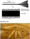
MK II sensor design (top) and use in hippocampal slices (bottom).
Fig. (6)
Fig. (6)
MK II sensor measurements of adenosine and ATP release during in vitro ischemia. A) Adenosine release increases gradually during the in vitro ischemic episode (black bar) then displays a surge on reoxygenation (black arrowhead). Short episodes of OGD elicit no ATP release. B) Longer episodes provoke the anoxic depolarisation and elicit ATP release, which also displays a surge on reoxygenation. Modified from [66].
Fig. (7)
Fig. (7)
Differential dependency of adenosine and ATP release on extracellular Ca2+. A) Release of adenosine during in vitro ischemia (black bar) is enhanced in nominally Ca2+-free aCSF, but ATP release is unaffected. B) Addition of EGTA to nominally Ca2+-free aCSF prevents ATP but not adenosine release. Modified from [66].
Fig. (8)
Fig. (8)
Release of adenosine during A) brief electrically-evoked seizures and B) spontaneous seizures in the presence of the A1R antagonist CPT. Periodic downward deflections reflect fEPSPs evoked at 15 s intervals. Following seizure activity the fEPSP is transiently depressed - less so in CPT, despite a much longer seizure. Modified from [55].

Similar articles

Cited by

References

    1. Abbracchio MP, Boeynaems JM, Barnard EA, Boyer JL, Kennedy C, Miras-Portugal MT, King BF, Gachet C, Jacobson KA, Weisman GA, Burnstock G. Characterization of the UDP-glucose receptor (re-named here the P2Y14 receptor) adds diversity to the P2Y receptor family. Trends Pharmacol. Sci. 2003;24:52–55. - PMC - PubMed
    1. Abbracchio MP, Burnstock G, Boeynaems JM, Barnard EA, Boyer JL, Kennedy C, Knight GE, Fumagalli M, Gachet C, Jacobson KA, Weisman GA. International Union of Pharmacology LVIII: update on the P2Y G protein-coupled nucleotide receptors: from molecular mechanisms and pathophysiology to therapy. Pharmacol. Rev. 2006;58:281–341. - PMC - PubMed
    1. Abbracchio MP, Burnstock G, Verkhratsky A, Zimmermann H. Purinergic signalling in the nervous system: an overview. Trends Neurosci. 2009;32:19–29. - PubMed
    1. Arrigoni E, Crocker AJ, Saper CB, Greene RW, Scammell TE. Deletion of presynaptic adenosine A1 receptors impairs the recovery of synaptic transmission after hypoxia. Neuroscience. 2005;132:575–580. - PMC - PubMed
    1. Bao L, Locovei S, Dahl G. Pannexin membrane channels are mechanosensitive conduits for ATP. FEBS Lett. 2004;572:65–68. - PubMed

LinkOut - more resources