

Adenosine and the auditory system
- PMID: 20190966
- PMCID: PMC2769008
- DOI: 10.2174/157015909789152155
Adenosine and the auditory system
Abstract
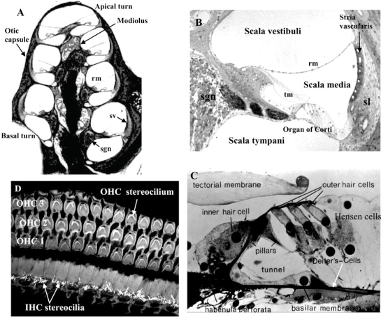
Adenosine is a signalling molecule that modulates cellular activity in the central nervous system and peripheral organs via four G protein-coupled receptors designated A(1), A(2A), A(2B), and A(3). This review surveys the literature on the role of adenosine in auditory function, particularly cochlear function and its protection from oxidative stress. The specific tissue distribution of adenosine receptors in the mammalian cochlea implicates adenosine signalling in sensory transduction and auditory neurotransmission although functional studies have demonstrated that adenosine stimulates cochlear blood flow, but does not alter the resting and sound-evoked auditory potentials. An interest in a potential otoprotective role for adenosine has recently evolved, fuelled by the capacity of A(1) adenosine receptors to prevent cochlear injury caused by acoustic trauma and ototoxic drugs. The balance between A(1) and A(2A) receptors is conceived as critical for cochlear response to oxidative stress, which is an underlying mechanism of the most common inner ear pathologies (e.g. noise-induced and age-related hearing loss, drug ototoxicity). Enzymes involved in adenosine metabolism, adenosine kinase and adenosine deaminase, are also emerging as attractive targets for controlling oxidative stress in the cochlea. Other possible targets include ectonucleotidases that generate adenosine from extracellular ATP, and nucleoside transporters, which regulate adenosine concentrations on both sides of the plasma membrane. Developments of selective adenosine receptor agonists and antagonists that can cross the blood-cochlea barrier are bolstering efforts to develop therapeutic interventions aimed at ameliorating cochlear injury. Manipulations of the adenosine signalling system thus hold significant promise in the therapeutic management of oxidative stress in the cochlea.
Keywords: Adenosine; adenosine receptors; cochlea; deafness; hearing; noise; ototoxicity.; oxidative stress.
Figures

References
-
- Adair TH. Growth regulation of the vascular system: an emerging role for adenosine. Am. J. Physiol. Regul. Integr. Comp. Physiol. 2005;289(2):R283–R296. - PubMed
-
- Alam SA, Oshima T, Suzuki M, Kawase T, Takasaka T, Ikeda K. The expression of apoptosis-related proteins in the aged cochlea of Mongolian gerbils. Laryngoscope. 2001;111:528–534. - PubMed
-
- Altschuler RA, Fairfield D, Cho Y, Leonova E, Benjamin IJ, Miller JM, Lomax MI. Stress pathways in the rat cochlea and potential for protection from acquired deafness. Audiol. Neurootol. 2002;7(3):152–156. - PubMed
-
- Baldwin SA, Mackey JR, Cass CE, Young JD. Nucleoside transporters: molecular biology and implications for therapeutic development. Mol. Med. Today. 1999;5:216–224. - PubMed
-
- Baldwin SA, Beal PR, Yao SY, King AE, Cass CE, Young JD. The equilibrative nucleoside transporter family, SLC29. Pflugers Arch. 2004;447(5):735–743. - PubMed
Grants and funding
LinkOut - more resources
Full Text Sources
Research Materials