

Ulnar nerve palsy following closed fracture of the distal radius: a report of 2 cases
- PMID: 20191002
- PMCID: PMC2824096
- DOI: 10.4055/cios.2010.2.1.55
Ulnar nerve palsy following closed fracture of the distal radius: a report of 2 cases
Abstract
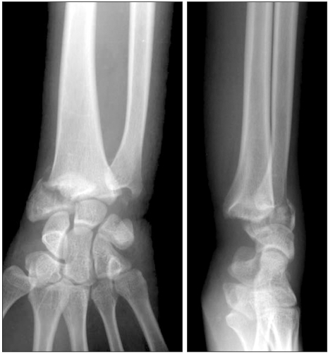
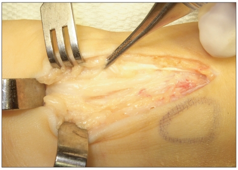
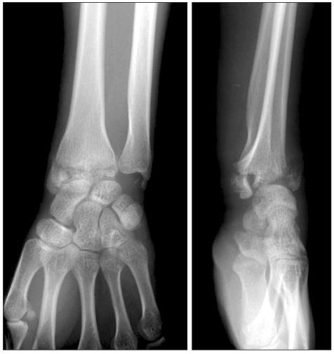
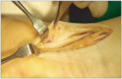
Ulnar nerve palsy subsequent to a fracture of the distal radius is extremely rare compared to a median nerve injury. The lesion tends to occur in younger patents with a high-energy mechanism of injury and a severe injury pattern consisting of wide displacement, comminution, combined distal ulnar fracture and open fracture. The mechanism of injury can contribute to a direct contusion and traction, compression secondary to prolonged edema and tissue fibrosis, intraneural fibrosis and laceration. We report 2 cases of progressive ulnar nerve palsy subsequent to closed fractures of the distal radius. The neurological symptoms recovered in all cases who underwent nerve decompression and neurolysis at 2 or 3 months after the trauma. It is recommended that cases with high-energy, widely displaced or comminuted fractures of the distal radius be evaluated carefully for ulnar nerve as well as median nerve injury.
Keywords: Closed; Distal; Palsy; Radius fracture; Ulnar nerve.
Figures
Similar articles
-
Dorsal displacement of the ulnar nerve after a displaced distal radius fracture: case report.J Hand Surg Am. 2009 Mar;34(3):432-5. doi: 10.1016/j.jhsa.2008.10.027. Epub 2009 Feb 12. J Hand Surg Am. 2009. PMID: 19216035
-
Combined median and ulnar nerve palsy complicating distal radius fractures.Orthop Traumatol Surg Res. 2018 Oct;104(6):871-875. doi: 10.1016/j.otsr.2018.04.026. Epub 2018 Jun 30. Orthop Traumatol Surg Res. 2018. PMID: 29969720
-
Ulnar nerve palsy associated with fracture of the distal radius.J Orthop Trauma. 2007 Feb;21(2):113-6. doi: 10.1097/BOT.0b013e31802f7335. J Orthop Trauma. 2007. PMID: 17304066
-
Ulnar nerve palsy following closed radiocarpal fracture-dislocation.Am J Orthop (Belle Mead NJ). 2008 Aug;37(8):E138-40. Am J Orthop (Belle Mead NJ). 2008. PMID: 18836611 Review. No abstract available.
-
Ulnar nerve laceration in a closed both bone forearm fracture.J Orthop Trauma. 1996;10(2):131-4. doi: 10.1097/00005131-199602000-00011. J Orthop Trauma. 1996. PMID: 8932674 Review.
Cited by
-
Ulnar nerve palsy as a complication of closed both-bone forearm fracture in a pediatric patient: a case report.Int Med Case Rep J. 2019 Mar 26;12:79-84. doi: 10.2147/IMCRJ.S200657. eCollection 2019. Int Med Case Rep J. 2019. PMID: 31114394 Free PMC article.
-
Ulnar Nerve and Ulnar Artery Injury Caused by Comminuted Distal Radius Fracture.J Orthop Case Rep. 2020 Jul;10(4):25-30. doi: 10.13107/jocr.2020.v10.i04.1786. J Orthop Case Rep. 2020. PMID: 33623761 Free PMC article.
-
Use of a novel acellular dermal matrix allograft to treat complex trauma wound: a case study.Int J Burns Trauma. 2014 Oct 26;4(2):62-5. eCollection 2014. Int J Burns Trauma. 2014. PMID: 25356372 Free PMC article.
-
Ulnar nerve injury associated with displaced distal radius fracture: Two case reports.World J Clin Cases. 2021 Aug 16;9(23):6956-6963. doi: 10.12998/wjcc.v9.i23.6956. World J Clin Cases. 2021. PMID: 34447848 Free PMC article.
-
Ulnar Nerve Translocation Following a Routine Distal Radius Fracture.Iowa Orthop J. 2023;43(1):185-189. Iowa Orthop J. 2023. PMID: 37383867 Free PMC article.
References
-
- Bourrel P, Ferro RM. Nerve complications in closed fractures of the lower end of the radius. Ann Chir Main. 1982;1(2):119–126. - PubMed
-
- Melone CP., Jr Articular fractures of the distal radius. Orthop Clin North Am. 1984;15(2):217–236. - PubMed
-
- Soong M, Ring D. Ulnar nerve palsy associated with fracture of the distal radius. J Orthop Trauma. 2007;21(2):113–116. - PubMed
-
- Bacorn RW, Kurtzke JF. Colles' fracture: a study of two thousand cases from the New York State Workmen's Compensation Board. J Bone Joint Surg Am. 1953;35(3):643–658. - PubMed
-
- Clarke AC, Spencer RF. Ulnar nerve palsy following fractures of the distal radius: clinical and anatomical studies. J Hand Surg Br. 1991;16(4):438–440. - PubMed
Publication types
MeSH terms
LinkOut - more resources
Full Text Sources
Medical
