

Case Reports
doi: 10.3348/kjr.2010.11.2.239.
Epub 2010 Feb 22.
Follicular dendritic cell sarcoma of the abdomen: the imaging findings
Affiliations
- PMID: 20191073
- PMCID: PMC2827789
- DOI: 10.3348/kjr.2010.11.2.239
Item in Clipboard
Case Reports
Follicular dendritic cell sarcoma of the abdomen: the imaging findings
Korean J Radiol.
2010 Mar-Apr.
Abstract
Follicular dendritic cell sarcoma is a rare neoplasm that originates from follicular dendritic cells in lymphoid follicles. This disease usually involves the lymph nodes, and especially the head and neck area. Rarely, extranodal sites may be affected, including tonsil, the oral cavity, liver, spleen and the gastrointestinal tract. We report here on the imaging findings of follicular dendritic cell sarcoma of the abdomen that involved the retroperitoneal lymph nodes and colon. It shows as a well-defined, enhancing homogenous mass with internal necrosis and regional lymphadenopathy.
Keywords: Abdomen, CT; Abdomen, US; Abdomen, neoplasms; Sarcoma.
Figures
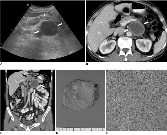
Follicular dendritic cell sarcoma in 69-year-old man. A. Abdominal ultrasonography shows 5 cm sized, well-defined anechoic mass (white arrows) that was located in posterior aspect of pancreas (P) with mild mass effect. Mass shows posterior acoustic enhancement that represents necrotic lesion. B. Axial CT scan shows well-defined necrotic mass with thin peripheral wall in retroperitoneal area (white arrows). Lesion shows mass effect on adjacent pancreas and splenic vein, but there was no evidence of direct invasion. C. Coronal reconstruction CT image shows several enlarged lymph nodes (black arrows) around cystic mass (white arrows). D. Gross specimen shows central whitish-yellow color that microscopically demonstrates extensive internal necrosis. E. Microscopic examination of solid component of mass reveals bundles of ovoid to spindle cells with pinkish cytoplasm (Hematoxylin & Eosin staining, ×100).
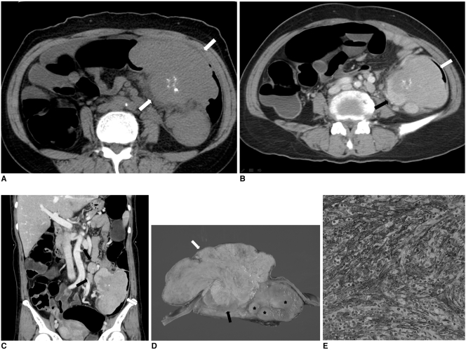
Follicular dendritic cell sarcoma in 52-year-old woman. A. Pre-contrast axial CT image shows multiple foci of irregular and dense calcifications in mass (white arrows). B. Post-contrast axial CT image shows well-defined large mass in mid-descending colon. Mass is composed of intraluminal (white arrow) and extraluminal (black arrow) components. Intraluminal component is less enhanced than extraluminal component. C. Coronal reconstruction CT image shows several enlarged regional lymph nodes (black arrows) around mass (white arrow). D. Gross specimen shows intraluminal component with relatively dull yellow color (white arrow) and non-necrotic area of extraluminal component (black arrow) with pink-tan color. Several enlarged pericolonic lymph nodes (asterisks) are noted. E. Immunohistochemical staining for CD35 demonstrates positive cytoplasmic expression (×400).
Similar articles
-
SIR 2008 annual meeting film panel case: Castleman disease complicated by follicular dendritic cell sarcoma.J Vasc Interv Radiol. 2008 Aug;19(8):1141-4. doi: 10.1016/j.jvir.2008.04.015. Epub 2008 Jun 6. J Vasc Interv Radiol. 2008. PMID: 18656005 No abstract available.
-
Computer Tomography Imaging Findings of Abdominal Follicular Dendritic Cell Sarcoma: A Report of 5 Cases.Medicine (Baltimore). 2016 Jan;95(1):e2404. doi: 10.1097/MD.0000000000002404. Medicine (Baltimore). 2016. PMID: 26735543 Free PMC article.
-
Follicular Dendritic Cell Sarcomas: CT and MRI Findings in 20 Patients.AJR Am J Roentgenol. 2021 Mar;216(3):835-843. doi: 10.2214/AJR.19.22759. Epub 2021 Jan 6. AJR Am J Roentgenol. 2021. PMID: 33405946
-
Clinicopathologic profile of intra-abdominal follicular dendritic cell sarcoma: A study of three cases with a literature review.Indian J Pathol Microbiol. 2024 Jan-Mar;67(1):195-200. doi: 10.4103/ijpm.ijpm_1089_21. Indian J Pathol Microbiol. 2024. PMID: 38358221 Review.
-
Follicular dendritic cell sarcoma arising from the lymph node of the pancreatic head: a case report with literature review.Clin J Gastroenterol. 2024 Aug;17(4):788-794. doi: 10.1007/s12328-024-01956-5. Epub 2024 Mar 26. Clin J Gastroenterol. 2024. PMID: 38532076 Review.
Cited by
-
Follicular dendritic cell sarcoma of the stomach in a young male: A rare case report and literature review.Medicine (Baltimore). 2025 Jul 4;104(27):e43004. doi: 10.1097/MD.0000000000043004. Medicine (Baltimore). 2025. PMID: 40629639 Free PMC article. Review.
-
Clinicopathological characteristics of mediastinal follicular dendritic cell sarcoma: report of three cases.J Cardiothorac Surg. 2016 Apr 11;11(1):56. doi: 10.1186/s13019-016-0464-5. J Cardiothorac Surg. 2016. PMID: 27068522 Free PMC article.
-
Follicular dendritic cell sarcoma of the omentum: multidetector computed tomography findings.Korean J Radiol. 2013 Mar-Apr;14(2):213-7. doi: 10.3348/kjr.2013.14.2.213. Epub 2013 Feb 22. Korean J Radiol. 2013. PMID: 23483068 Free PMC article.
-
Follicular dendritic cell sarcoma of gastrointestinal tract with two emerging distinct subtypes: a case report and systemic review.Diagn Pathol. 2022 Aug 8;17(1):64. doi: 10.1186/s13000-022-01246-z. Diagn Pathol. 2022. PMID: 35941667 Free PMC article.
-
Imaging findings of follicular dendritic cell sarcoma: report of four cases.Korean J Radiol. 2011 Jan-Feb;12(1):122-8. doi: 10.3348/kjr.2011.12.1.122. Epub 2011 Jan 3. Korean J Radiol. 2011. PMID: 21228948 Free PMC article.
References
-
- Tew JG, Kosco MH, Burton GF, Szakal AK. Follicular dendritic cells as accessory cells. Immunol Rev. 1990;117:185–211. - PubMed
-
- Chan JK, Banks PM, Cleary ML, Delsol G, De Wolf-Peeters C, Falini B, et al. A revised European-American classification of lymphoid neoplasms proposed by the International Lymphoma Study Group. A summary version. Am J Clin Pathol. 1995;103:543–560. - PubMed
-
- Chan JK, Fletcher CD, Nayler SJ, Cooper K. Follicular dendritic cell sarcoma. Clinicopathologic analysis of 17 cases suggesting a malignant potential higher than currently recognized. Cancer. 1997;79:294–313. - PubMed
-
- Kairouz S, Hashash J, Kabbara W, McHayleh W, Tabbara IA. Dendritic cell neoplasms: an overview. Am J Hematol. 2007;82:924–928. - PubMed
Publication types
MeSH terms
LinkOut - more resources
Full Text Sources
