

Measurement of isoprostanes as markers of oxidative stress in neuronal tissue
- PMID: 20191108
- PMCID: PMC2828759
- DOI: 10.1002/0471140856.tx1214s39
Measurement of isoprostanes as markers of oxidative stress in neuronal tissue
Abstract
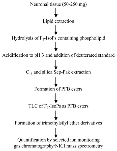
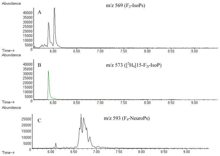
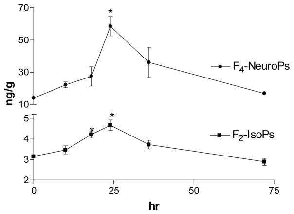
Oxidative stress is implicated in the pathogenesis of a variety of human diseases, including neurodegenerative disease, atherosclerosis and cancer, as well as progressive and even normal aging processes. Increased generation of free radicals derived primarily from molecular oxygen has also been associated with neuronal damage induced by a variety of environmental agents. However, measuring oxidative stress in biological systems is complex and requires accurate quantification of either free radicals or damaged biomolecules. One method to quantify oxidative injury is to measure lipid peroxidation. Lipids are readily attacked by free radicals, resulting in the formation of a number of peroxidation products. F₂-isoprostanes (F₂-IsoPs) are one group of these compounds, which are derived by the free radical peroxidation of arachidonic acid (AA). The F₂-IsoPs, prostaglandine F₂-like compounds, have been shown as the most accurate measure of oxidative damage in vivo. This review summarizes current methodology used to quantify F₂-IsoPs and discusses the utility of these and other prostaglandine (PG)-like compounds as in vivo biomarkers of oxidative stress in neuronal tissues.
Keywords: F2-isoprostanes; lipid peroxidation; neuroprostanes; oxidative damage.
Figures
Similar articles
-
Measurement of isoprostanes as markers of oxidative stress.Methods Mol Biol. 2011;758:195-204. doi: 10.1007/978-1-61779-170-3_13. Methods Mol Biol. 2011. PMID: 21815067 Free PMC article.
-
Insights into oxidative stress: the isoprostanes.Curr Med Chem. 2007;14(6):703-17. doi: 10.2174/092986707780059607. Curr Med Chem. 2007. PMID: 17346157 Review.
-
Quantification of F2-isoprostanes in biological fluids and tissues as a measure of oxidant stress.Methods Enzymol. 2007;433:113-26. doi: 10.1016/S0076-6879(07)33006-1. Methods Enzymol. 2007. PMID: 17954231
-
F2-isoprostanes biomarkers of lipid peroxidation: their utility in evaluation of oxidative stress induced by toxic agents.Int J Occup Med Environ Health. 2002;15(1):19-27. Int J Occup Med Environ Health. 2002. PMID: 12038860 Review.
-
Measurement of products of docosahexaenoic acid peroxidation, neuroprostanes, and neurofurans.Methods Enzymol. 2007;433:127-43. doi: 10.1016/S0076-6879(07)33007-3. Methods Enzymol. 2007. PMID: 17954232
Cited by
-
Prostanoid signaling: dual role for prostaglandin E2 in neurotoxicity.Neurotoxicology. 2011 Jun;32(3):312-9. doi: 10.1016/j.neuro.2011.02.004. Epub 2011 Mar 3. Neurotoxicology. 2011. PMID: 21376752 Free PMC article. Review.
-
Chlorpyrifos-, diisopropylphosphorofluoridate-, and parathion-induced behavioral and oxidative stress effects: are they mediated by analogous mechanisms of action?Toxicol Sci. 2013 Jan;131(1):206-16. doi: 10.1093/toxsci/kfs280. Epub 2012 Sep 17. Toxicol Sci. 2013. PMID: 22986948 Free PMC article.
-
Isoprostanes and neuroprostanes as biomarkers of oxidative stress in neurodegenerative diseases.Oxid Med Cell Longev. 2014;2014:572491. doi: 10.1155/2014/572491. Epub 2014 Apr 29. Oxid Med Cell Longev. 2014. PMID: 24868314 Free PMC article. Review.
-
Potential Correlation Between Molecular Biomarkers and Oxidative Stress in Traumatic Brain Injury.Int J Mol Sci. 2025 Apr 18;26(8):3858. doi: 10.3390/ijms26083858. Int J Mol Sci. 2025. PMID: 40332547 Free PMC article. Review.
-
Tissue pretreatment for LC-MS/MS analysis of PUFA and eicosanoid distribution in mouse brain and liver.Anal Bioanal Chem. 2020 Apr;412(10):2211-2223. doi: 10.1007/s00216-019-02170-w. Epub 2019 Dec 21. Anal Bioanal Chem. 2020. PMID: 31865417 Free PMC article.
References
-
- Basu S. Radioimmunoassay of 8-iso-prostaglandin F2alpha: an index of oxidative injury via free radical catalysed lipid peroxidation. Prostaglandins Leukot Essent Fatty Acids. 1998;58:319–325. - PubMed
-
- Basu S. F2-isoprostanes in human health and diseases: from molecular mechanisms to clinical implications. Antioxidants and Redox Signaling. 2008;10:1405–1434. - PubMed
-
- Ben-Ari Y, Cossart R. Kainate, a double agent that generates seizures: two decades of progress. Trends Neurosci. 2000;23:580–587. - PubMed
-
- Famm SS, Morrow JD. The isoprostanes: Unique products of arachidonic acid oxidation- a review. Curr Med Chem. 2003;10:1723–1740. - PubMed
Publication types
MeSH terms
Substances
Grants and funding
LinkOut - more resources
Full Text Sources
Miscellaneous
