

Impact of analyzing less image frames per segment for radiofrequency-based volumetric intravascular ultrasound measurements in mild-to-moderate coronary atherosclerosis
- PMID: 20191323
- PMCID: PMC2868170
- DOI: 10.1007/s10554-010-9599-y
Impact of analyzing less image frames per segment for radiofrequency-based volumetric intravascular ultrasound measurements in mild-to-moderate coronary atherosclerosis
Abstract
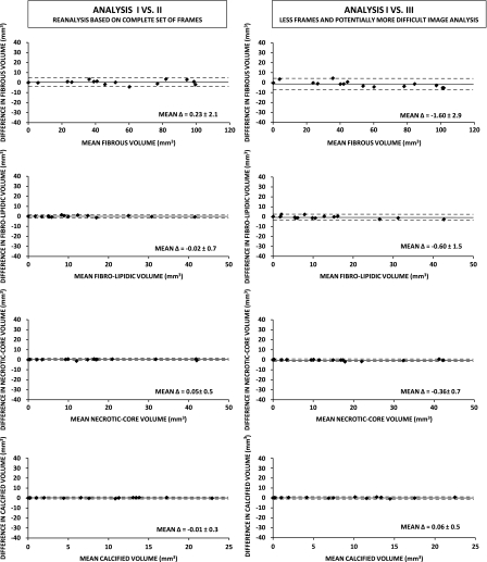
Volumetric radiofrequency-based intravascular ultrasound (RF-IVUS) data of coronary segments are increasingly used as endpoints in serial trials of novel anti-atherosclerotic therapies. In a relatively time-consuming process, vessel and lumen contours are defined; these contours are first automatically detected, then visually checked, and finally (in most cases) manually edited to generate reliable volumetric data of vessel geometry and plaque composition. Reduction in number of cross-sectional images for volumetric analysis could save analysis time but may also increase measurement variability of volumetric data. To assess whether a 50% reduction in number of frames per segment (every second frame) alters the reproducibility of volumetric measurements, we performed repeated RF-IVUS analyses of 15 coronary segments with mild-to-moderate atherosclerosis (20.2 +/- 0.2 mm-long segments with 46 +/- 13% plaque burden). Volumes were calculated based on a total of 731 image frames. Reducing the number of cross-sectional image frames for volumetric measurements saved analysis time (38 +/- 9 vs. 68 +/- 17 min/segment; P < 0.0001) and resulted for only a few parameters in (borderline) significant but mild differences versus measurements based on all frames (fibrous volume, P < 0.05; necrotic-core volume, P = 0.07). Compared to the intra-observer variability, there was a mild increase in measurement variability for most geometrical and compositional volumetric RF-IVUS parameters. In RF-IVUS studies of mild-to-moderate coronary disease, analyzing less image frames saved analysis time, left most volumetric parameters greatly unaffected, and resulted in a no more than mild increase in measurement variability of volumetric data.
Figures


Similar articles
-
Impact of analyzing fewer image frames per segment during offline volumetric radiofrequency-based intravascular ultrasound measurements of target lesions prior to percutaneous coronary interventions.Int J Cardiovasc Imaging. 2012 Mar;28(3):479-89. doi: 10.1007/s10554-011-9843-0. Epub 2011 Mar 19. Int J Cardiovasc Imaging. 2012. PMID: 21424153 Free PMC article.
-
Reproducibility of volumetric intravascular ultrasound radiofrequency-based analysis of coronary plaque composition in vivo.Int J Cardiovasc Imaging. 2009 Jan;25(1):13-23. doi: 10.1007/s10554-008-9338-9. Epub 2008 Aug 13. Int J Cardiovasc Imaging. 2009. PMID: 18704753 Free PMC article.
-
Reproducibility of Shin's method for necrotic core and calcium content in atherosclerotic coronary lesions treated with bioresorbable everolimus-eluting vascular scaffolds using volumetric intravascular ultrasound radiofrequency-based analysis.Int J Cardiovasc Imaging. 2012 Jan;28(1):43-9. doi: 10.1007/s10554-010-9779-9. Epub 2011 Jan 13. Int J Cardiovasc Imaging. 2012. PMID: 21229393
-
Intravascular ultrasound in the coronary arteries.Semin Vasc Surg. 2006 Sep;19(3):132-8. doi: 10.1053/j.semvascsurg.2006.06.007. Semin Vasc Surg. 2006. PMID: 16996414 Review.
-
Quantitative three-dimensional intravascular ultrasound.Semin Interv Cardiol. 1997 Mar;2(1):25-32. Semin Interv Cardiol. 1997. PMID: 9546981 Review.
Cited by
-
Ultrasound and light: friend or foe? On the role of intravascular ultrasound in the era of optical coherence tomography.Int J Cardiovasc Imaging. 2011 Feb;27(2):209-14. doi: 10.1007/s10554-011-9797-2. Epub 2011 Feb 20. Int J Cardiovasc Imaging. 2011. PMID: 21337025 Free PMC article.
-
Impact of analyzing fewer image frames per segment during offline volumetric radiofrequency-based intravascular ultrasound measurements of target lesions prior to percutaneous coronary interventions.Int J Cardiovasc Imaging. 2012 Mar;28(3):479-89. doi: 10.1007/s10554-011-9843-0. Epub 2011 Mar 19. Int J Cardiovasc Imaging. 2012. PMID: 21424153 Free PMC article.
-
Multicenter assessment of the reproducibility of volumetric radiofrequency-based intravascular ultrasound measurements in coronary lesions that were consecutively stented.Int J Cardiovasc Imaging. 2012 Dec;28(8):1867-78. doi: 10.1007/s10554-012-0011-y. Epub 2012 Jan 14. Int J Cardiovasc Imaging. 2012. PMID: 22246064 Free PMC article.
References
-
- Bruining N, von Birgelen C, de Feyter PJ, Ligthart J, Li W, Serruys PW, Roelandt JR. ECG-gated versus nongated three-dimensional intracoronary ultrasound analysis: implications for volumetric measurements. Cathet Cardiovasc Diagn. 1998;43(3):254–260. doi: 10.1002/(SICI)1097-0304(199803)43:3<254::AID-CCD3>3.0.CO;2-8. - DOI - PubMed
-
- Mintz GS, Nissen SE, Anderson WD, Bailey SR, Erbel R, Fitzgerald PJ, Pinto FJ, Rosenfield K, Siegel RJ, Tuzcu EM, Yock PG. American college of cardiology clinical expert consensus document on standards for acquisition, measurement and reporting of intravascular ultrasound studies (IVUS). A report of the American college of cardiology task force on clinical expert consensus documents. J Am Coll Cardiol. 2001;37(5):1478–1492. doi: 10.1016/S0735-1097(01)01175-5. - DOI - PubMed
-
- von Birgelen C, de Vrey EA, Mintz GS, Nicosia A, Bruining N, Li W, Slager CJ, Roelandt JR, Serruys PW, de Feyter PJ. ECG-gated three-dimensional intravascular ultrasound: feasibility and reproducibility of the automated analysis of coronary lumen and atherosclerotic plaque dimensions in humans. Circulation. 1997;96(9):2944–2952. - PubMed
MeSH terms
LinkOut - more resources
Full Text Sources
Medical
