. 2010 Apr 15;82(8):3118-23.
doi: 10.1021/ac902802b.

Ultrasensitive electrochemical immunosensor for oral cancer biomarker IL-6 using carbon nanotube forest electrodes and multilabel amplification

Affiliations

Ultrasensitive electrochemical immunosensor for oral cancer biomarker IL-6 using carbon nanotube forest electrodes and multilabel amplification

Ruchika Malhotra et al. Anal Chem. .

Abstract

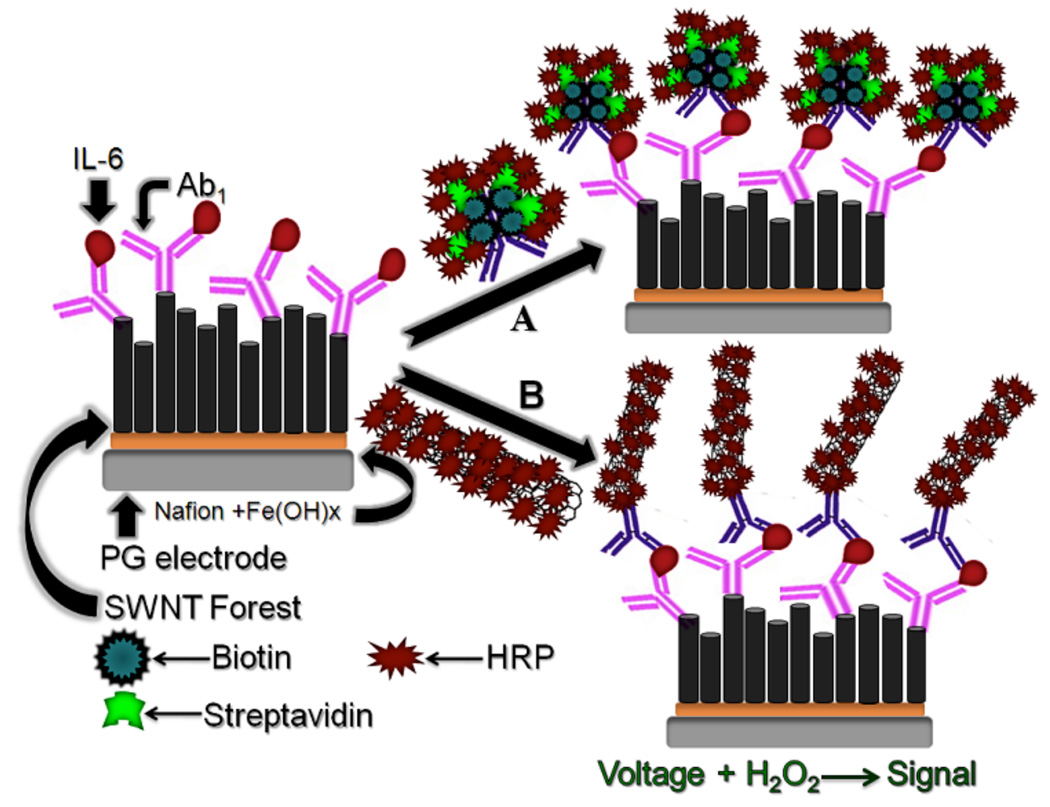
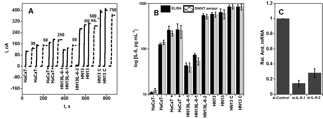
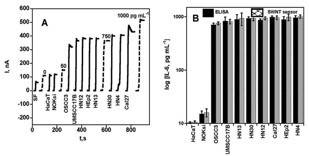
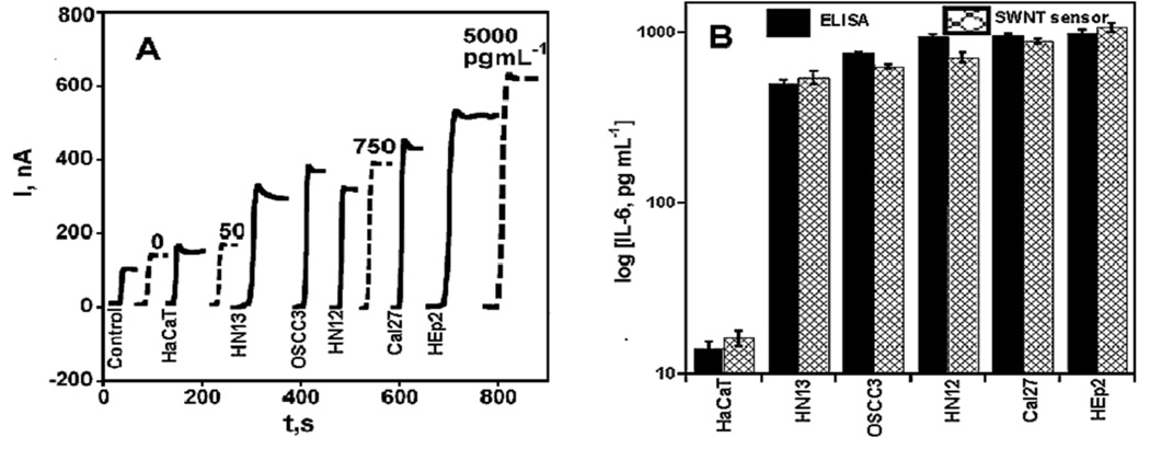
Squamous cell carcinomas of head and neck (HNSCC) are associated with immune, inflammatory, and angiogenic responses involving interleukin-6 (IL-6). This article reports an ultrasensitive electrochemical immunosensor for human IL-6 and proof-of-concept studies of IL-6 detection in HNSCC cells. Single wall carbon nanotube (SWNT) forests with attached capture antibodies (Ab(1)) for IL-6 were used in an electrochemical sandwich immunoassay protocol using enzyme label horseradish peroxidase (HRP) to measure very low (<or=30 pg mL(-1)) and elevated levels of IL-6. Two levels of multienzyme labeling were used to measure a broad concentration range of IL-6 in a representative panel of HNSCC cells. Secondary antibodies (Ab(2)) attached to carboxylated multiwall carbon nanotubes with 106 HRP labels per 100 nm gave the highest sensitivity of 19.3 nA mL (pg IL-6)(-1) cm(-2) and the best detection limit (DL) of 0.5 pg mL(-1) (25 fM) for IL-6 in 10 microL of calf serum. For more concentrated samples, biotinylated Ab(2) bound to streptavidin-HRP to provide 14-16 labels per antigen was used. These immunosensors accurately measured secreted IL-6 in a wide range of HNSCC cells demonstrated by excellent correlations with standard enzyme-linked immunosorbent assays (ELISA), suggesting that SWNT immunosensors combined with multilabel detection have excellent promise for detecting IL-6 in research and clinical applications.


Figures

Figure 1
Figure 1
Two strategies for multilabel detection in amperometric immunosensor (A) immunosensor after treating with Ab2-biotin-streptavidin-HRP, providing 14 to 16 HRP’s on one Ab2; (B) immunosensor after treating with HRP-MWNT-Ab2 bioconjugate having 106 active HRPs (enzyme labels) per 100 nm of carboxylated carbon nanotubes. The final detection step involves immersing the immunosensor in an electrochemical cell containing PBS buffer and mediator, applying voltage and injecting a small amount of hydrogen peroxide.
Figure 2
Figure 2
Amperometric response for SWNT immunosensors incubated with human IL-6 in 10 µL calf serum, then biotin-Ab2-MWNT-HRP (A) current at −0.3 V and rotating the electrode at 3000 rpm in PBS buffer containing 1 mM hydroquinone mediator and then injecting H2O2 to 0.04 mM to develop the signal; (B) influence of IL-6 concentration on steady-state current (corrected for background) for immunosensor using the bioconjugate. Error bars represent device-to-device standard deviation (n = 3).
Figure 3
Figure 3
Amperometric response for SWNT immunosensors incubated with IL-6 in 10 µL calf serum (pg mL−1 labeled on curves, dashed lines) or conditioned media from cells treated with TNF-alpha (HaCaT −/+) and those transfected with indicated siRNA oligonucleotides (HN13IL-6-1, HN13IL-6-2, HN13 [non transfected] and HN13 C [control siRNA]; solid lines) for 1 h. Conditioned media samples (HaCaT−, HaCaT+, HN13IL-6-1, HN13IL-6-2, HN13, HN13 C) were analyzed using 10 µL 1.1 pmol L−1 biotinylated secondary antibody (Ab2) in 0.1 % BSA in pH 7.2 PBS buffer and 10 µL streptavidin-HRP except the one below 30 pg mL−1 (HaCaT−), which was analyzed using 10 µL Ab2-MWNT-HRP bioconjugates (A) current at −0.3 V and 3000 rpm using 1 mM hydroquinone as mediator in PBS buffer, then injecting H2O2 to 0.4 mM; (B) SWNT sensor results for conditioned media shown with results from ELISA for the same samples; (C) qRT-PCR analysis of RNA extracted from control and IL-6 siRNA transfected cells and expression levels indicated (fold) are after normalization to levels of GAPDH.
Figure 4
Figure 4
Amperometric response for SWNT immunosensors incubated with IL-6 in 10 µL calf serum (pg mL−1 labeled on curves, dashed lines) and conditioned media (HaCaT, Cal27, HEp2, HN4, HN12, HN13, HN30, NOKsi, OSCC3, UMSCC17B) and serum free media, SF (solid lines) for 1 h. Conditioned media samples (Cal27, HEp2, HN4, HN12, HN13, HN30, OSCC3, UMSCC17B) were analyzed using 10 µL 1.1 pmol L−1 biotinylated secondary antibody (Ab2) in 0.1 % BSA in pH 7.2 PBS buffer and 10 µL streptavidin-HRP except those below 30 pg mL−1 (HaCaT, NOKsi), which were analyzed using 10 µL Ab2-MWNT-HRP bioconjugates (A) current at −0.3 V and 3000 rpm using 1 mM hydroquinone as mediator in PBS buffer, then injecting H2O2 to 0.4 mM; (B) SWNT sensor results for conditioned media shown with results from ELISA (relative standard deviation [ RSD] ± 10%) for the same samples.
Figure 5
Figure 5
Amperometric response for SWNT immunosensors incubated with IL-6 in 10 µL calf serum (pg mL−1 labeled on curves, dashed lines) and a second set of conditioned media (Cal27, HaCaT, HEp2, HN12, HN13, OSCC3) and control (solid lines) for 1 h. Conditioned media samples (Cal27, HEp2, HN12, HN13, OSCC3) were analyzed using 10 µL 1.1 pmol L−1 biotinylated secondary antibody (Ab2) in 0.1 % BSA in pH 7.2 PBS buffer and 10 µL streptavidin-HRP except the one below 30 pg mL−1 (HaCaT), which was analyzed using 10 µL Ab2-MWNT-HRP bioconjugates (A) current at −0.3 V and 3000 rpm using 1 mM hydroquinone as mediator in PBS buffer, then injecting H2O2 to 0.4 mM; (B) SWNT sensor results for conditioned media shown with results from ELISA (RSD ± 10%) for the same samples.

Similar articles

Cited by

References

    1. Kishimoto T. Annu. Rev. Immunol. 2005;23:1–21. - PubMed
    1. Jemal A, Murray T, Ward E. CA Cancer J. Clin. 2005;55:10–30. - PubMed
    1. Thomas GR, Nadiminti H, Regalado J. Int. J. Exp. Pathol. 2005;86:347–363. - PMC - PubMed
    1. Riedel F, Zaiss I, Herzog D, Götte K, Naim R, Hörmann K. Anticancer Res. 2005;25:2761–2765. - PubMed
    1. Hong DS, Angelo LS, Kurzrock R. Cancer. 2007;110:1911–1928. - PubMed

Publication types