Review
. 2010:39:329-48.
doi: 10.1146/annurev.biophys.093008.131400.

Optical control of neuronal activity

Affiliations
Review

Optical control of neuronal activity

Stephanie Szobota et al. Annu Rev Biophys. 2010.

Abstract

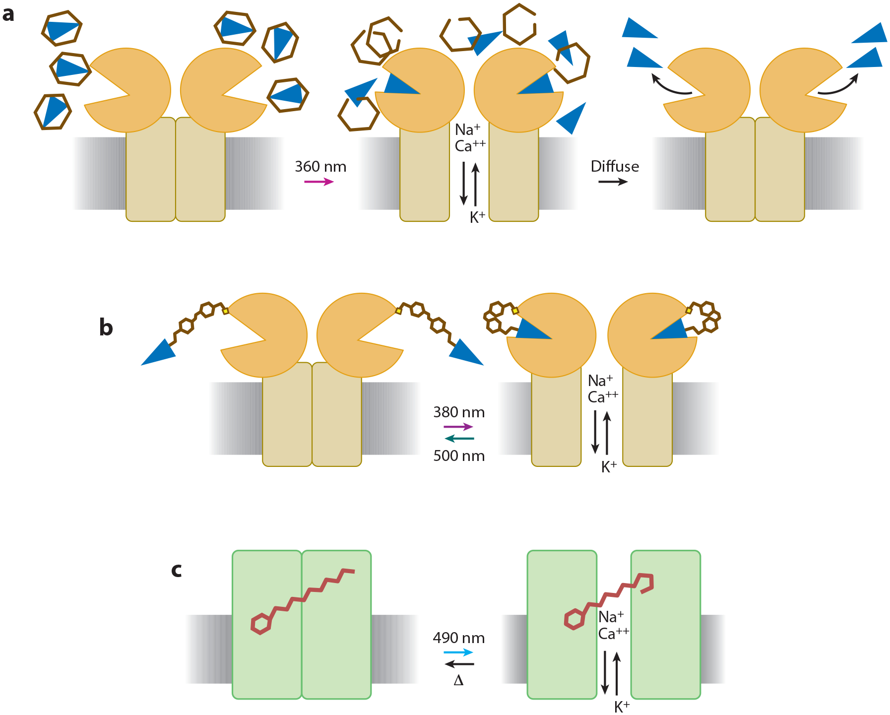
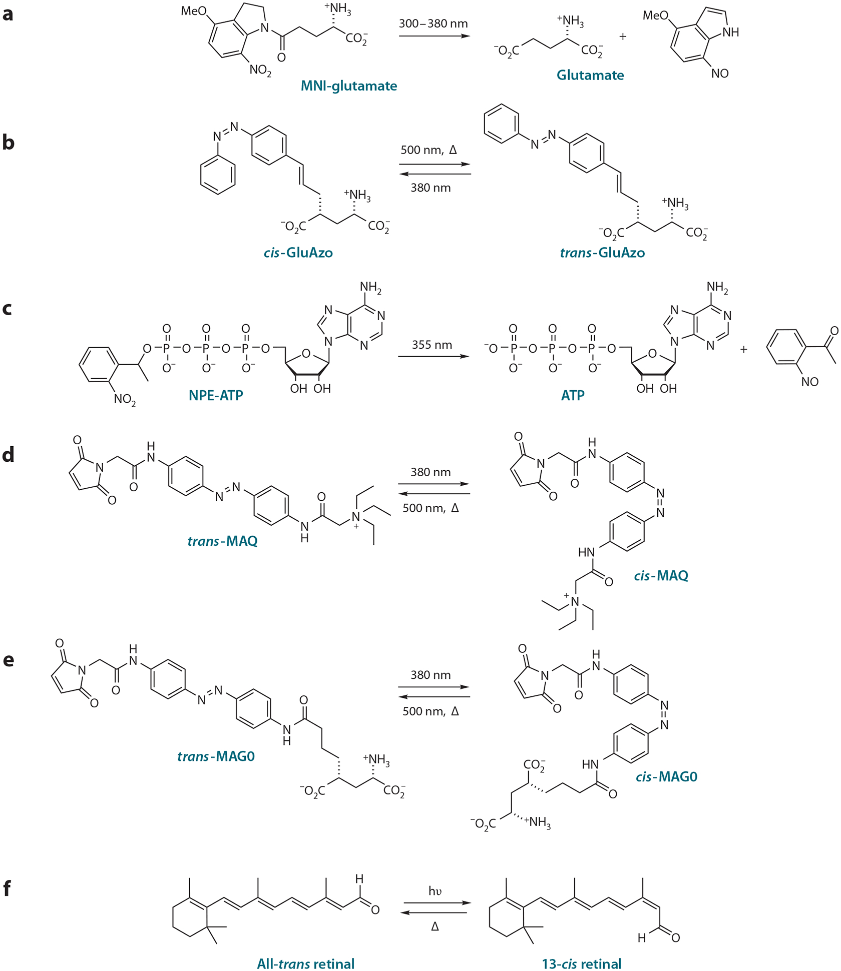
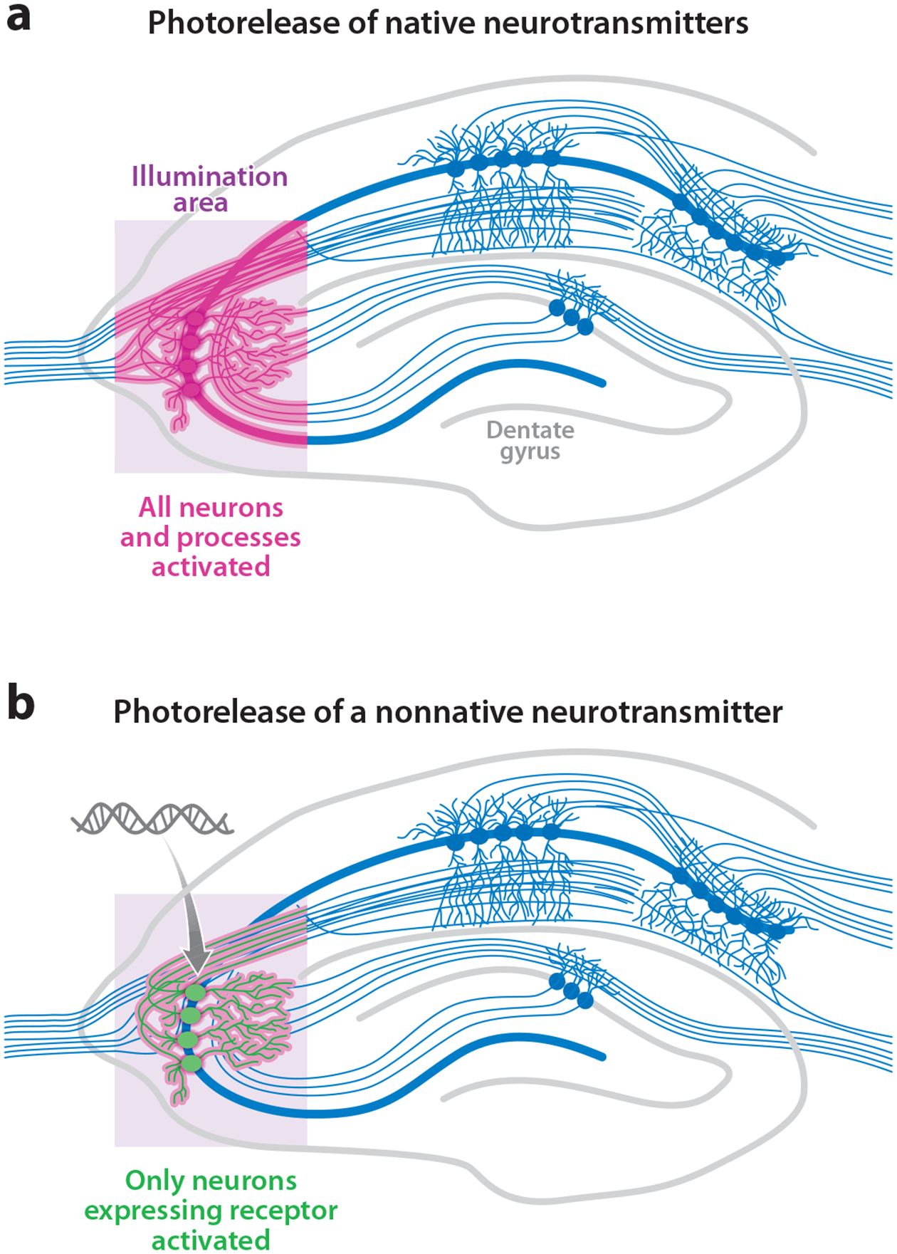
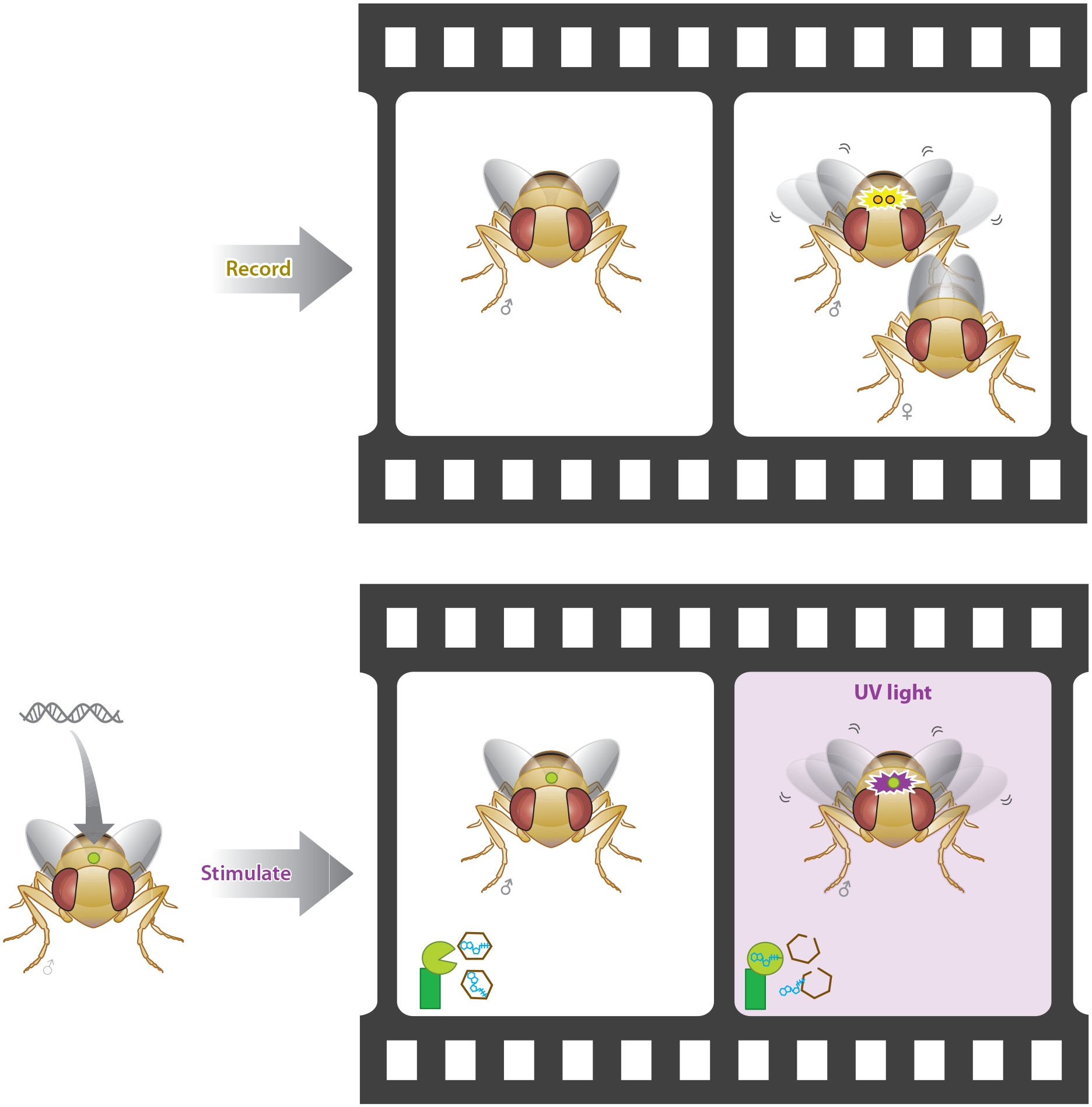
Advances in optics, genetics, and chemistry have enabled the investigation of brain function at all levels, from intracellular signals to single synapses, whole cells, circuits, and behavior. Until recent years, these research tools have been utilized in an observational capacity: imaging neural activity with fluorescent reporters, for example, or correlating aberrant neural activity with loss-of-function and gain-of-function pharmacological or genetic manipulations. However, optics, genetics, and chemistry have now combined to yield a new strategy: using light to drive and halt neuronal activity with molecular specificity and millisecond precision. Photostimulation of neurons is noninvasive, has unmatched spatial and temporal resolution, and can be targeted to specific classes of neurons. The optical methods developed to date encompass a broad array of strategies, including photorelease of caged neurotransmitters, engineered light-gated receptors and channels, and naturally light-sensitive ion channels and pumps. In this review, we describe photostimulation methods, their applications, and opportunities for further advancement.


Figures

Figure 1
Figure 1
Photostimulation technology can be classified into three general categories. (a) Caged compounds can be released by a flash of light, allowing the liberated compound to act on endogenous or exogenous neuronal targets before diffusing away. (b) Neurotransmitter receptors can be engineered to become light sensitive when coupled to a synthetic photoswitchable molecule containing an analogue of the native neurotransmitter. (c) Naturally light-sensitive proteins can be expressed in neurons; these proteins interact with the chromophore retinal, which is native to the nervous system of many animals, to photoregulate membrane potential.
Figure 2
Figure 2
Light-sensitive compounds enable photostimulation of neurons. (a) Caged glutamate acts on native glutamate receptors after it is uncaged by a pulse of UV light. (b) GluAzo is a reversibly caged neurotransmitter acting on native glutamate receptors when photoisomerized to the active trans configuration. (c) Caged ATP acts on nonnative P2X2 receptors that can be expressed in invertebrate neurons that lack ATP receptors and do not use ATP as a transmitter. (d) The cis form of maleimide-azobenzene-quaternary ammonium (MAQ), when coupled to an engineered Shaker K+ channel, silences neuronal activity by unblocking the pore, a process that is reversed when photoisomerized to the trans form. (e) Maleimide-azobenzene-glutamate (MAG) attaches covalently to an introduced cysteine in glutamate receptors and activates them when it is photoisomerized to the active isoform. Depending on the attachment site, activation is by either cis or trans MAG. MAG0 is the shortest of three different versions of MAG. (f) Retinal-coupled proteins like channelrhodopsin and halorhodopsin become activated when all-trans retinal is converted to 13-cis retinal (at wavelengths dictated by the specific interactions between the chromophore and protein) and then relax to the inactive state in the dark. We thank Matthew Volgraf for drawing the chemical structures.
Figure 3
Figure 3
Photostimulation of neurons has additional genetic resolution when neurons are induced to be light sensitive by genetic targeting. (a) Photorelease of native neurotransmitters activates all the neurons and neuronal processes within the illumination region. (b) Photorelease of a nonnative transmitter activates only the neurons within the illumination region that are also expressing the exogenous transmitter receptor. This genetic specificity also exists for opsins and photoswitched tethered ligand-gated channels. Image modified after http://www.bris.ac.uk/Depts/Synaptic/info/pathway/figs/hippocampus.gif
Figure 4
Figure 4
Genetic targeting of specific neural circuits enables optical gating of natural behaviors. In this example, generation of a courtship song involves wing vibrations and a complex behavior that engages many neural circuits. This neural activity can be recorded, and a subset of those neurons can then be stimulated to establish causation of the behavior using optogenetic stimulation (22).
Figure 5
Figure 5
Optogenetic strategies for photostimulation. (a) A fly learns that an odor is aversive when the odor is paired with optical activation of P2X2 receptors in circumscribed dopaminergic neurons (21). (b) A zebrafish engages in a directed C-turn escape maneuver upon optical stimulation of LiGluR in defined sensory neurons (89). (c) A mouse wakes up when hypocretin neurons in the hypothalamus expressing channelrhodopsin are optically stimulated (1).
Figure 6
Figure 6
Some photostimulation methods allow activity to persist in the dark, enabling observation of behaviors that would otherwise be perturbed by constant illumination. (a) LiGluR remains activated (for tens of minutes) after brief exposure to ~380-nm light (34). The same is true of some ChR2 mutants, though for shorter times (12). (b) A hippocampal neuron expressing LiGluR fires action potentials when illuminated at 374 nm for 5 ms and then continues to fire in the dark until 488-nm light restores native activity. (c) Zebrafish naturally respond to a mechanical poke in the side by making a directed C-turn to escape the stimulus. (d) Illumination at ~380 nm followed by a long period of darkness induces a numbing of the escape reflex, which is then restored after exposure to ~500-nm light (79). (e) A focused optical poke with high-intensity 380 nm prevents light from entering the eyes and induces the same C-turn escape maneuver as in panel c (89).

References

    1. Adamantidis AR, Zhang F, Aravanis AM, Deisseroth K, de Lecea L. 2007. Neural substrates of awakening probed with optogenetic control of hypocretin neurons. Nature 450(7168):420–24 - PMC - PubMed
    1. Ahmed NA, Radwan NM, Ibrahim KM, Khedr ME, El Aziz MA, Khadrawy YA. 2008. Effect of three different intensities of infrared laser energy on the levels of amino acid neurotransmitters in the cortex and hippocampus of rat brain. Photomed. Laser Surg 26(5):479–88 - PubMed
    1. Airan RD, Thompson KR, Fenno LE, Bernstein H, Deisseroth K. 2009. Temporally precise in vivo control of intracellular signalling. Nature 458(7241):1025–29 - PubMed
    1. Alilain WJ, Li X, Horn KP, Dhingra R, Dick TE, et al. 2008. Light-induced rescue of breathing after spinal cord injury. J. Neurosci 28(46):11862–70 - PMC - PubMed
    1. Allégre G, Avrillier S, Albe-Fessard D. 1994. Stimulation in the rat of a nerve fiber bundle by a short UV pulse from an excimer laser. Neurosci. Lett 180(2):261–64 - PubMed

Publication types

LinkOut - more resources