Review
. 2010:39:265-89.
doi: 10.1146/annurev.biophys.093008.131228.

Eukaryotic chemotaxis: a network of signaling pathways controls motility, directional sensing, and polarity

Affiliations
Review

Eukaryotic chemotaxis: a network of signaling pathways controls motility, directional sensing, and polarity

Kristen F Swaney et al. Annu Rev Biophys. 2010.

Abstract

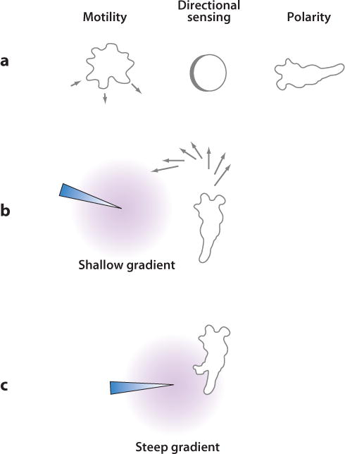
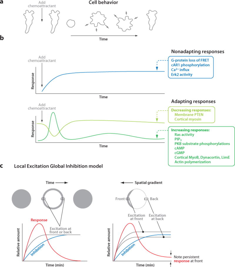
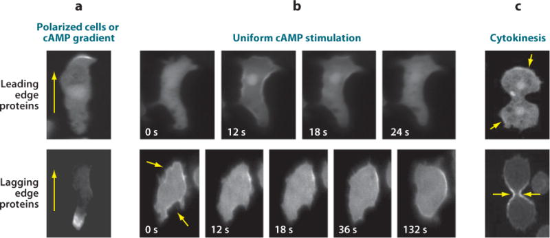
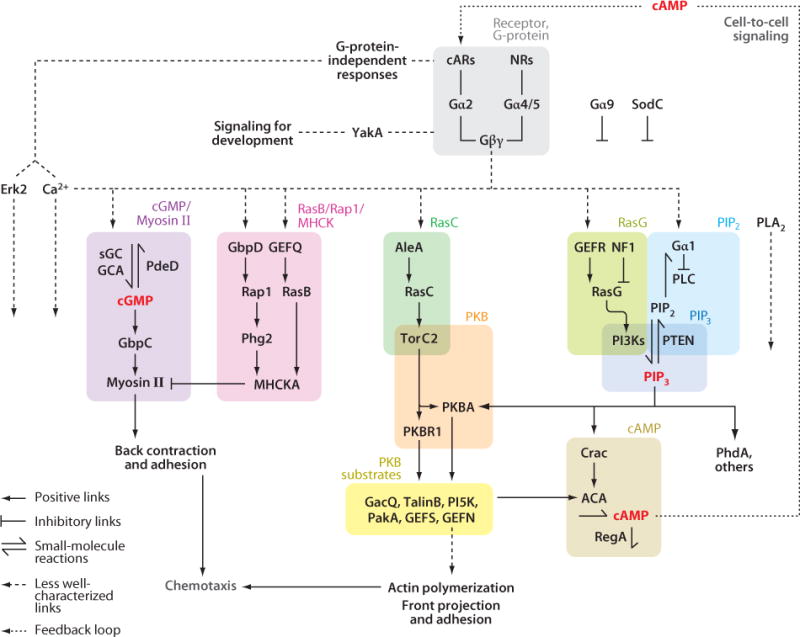
Chemotaxis, the directed migration of cells in chemical gradients, is a vital process in normal physiology and in the pathogenesis of many diseases. Chemotactic cells display motility, directional sensing, and polarity. Motility refers to the random extension of pseudopodia, which may be driven by spontaneous actin waves that propagate through the cytoskeleton. Directional sensing is mediated by a system that detects temporal and spatial stimuli and biases motility toward the gradient. Polarity gives cells morphologically and functionally distinct leading and lagging edges by relocating proteins or their activities selectively to the poles. By exploiting the genetic advantages of Dictyostelium, investigators are working out the complex network of interactions between the proteins that have been implicated in the chemotactic processes of motility, directional sensing, and polarity.


Figures

Figure 1
Figure 1
Chemotaxis is composed of motility, polarity, and directional sensing. In the presence of a chemoattractant (or chemorepellent) gradient, cells move toward (or away from) higher concentrations. (a) Left: Free amoeboid cells rhythmically extend pseudopodia and move in random directions. Middle: Spatial sensing, a means of directional sensing, can be demonstrated by the gradient-mediated relocalization of proteins in cells immobilized by actin inhibitors. Right: Chemotactic cells are often polarized, with a stable leading edge from which pseudopodia are extended. (b) In a shallow gradient, polarized cells display biased patterns of pseudopodia extension at the leading edge that cause cells to turn gradually toward higher concentrations of chemoattractant. (c) Sufficiently steep gradients can trigger new projections anywhere along the cell periphery.
Figure 2
Figure 2
Temporal and spatial responses triggered by chemoattractants and the Local Excitation Global Inhibition (LEGI) model. (a) When exposed to a sudden increase in cAMP, cells retract projections or cringe; then, they periodically extend and retract projections at random sites on the periphery until, after several minutes, they regain their polarized morphology. (b) The biochemical responses triggered by cAMP can be divided into two groups on the basis of whether or not they adapt to constant stimuli. Some responses, such as G-protein activation, are nonadapting and persist as long as the stimuli are maintained. Of the adapting responses, most, such as PIP3 production, transiently increase, whereas others, such as membrane-localized PTEN, transiently decrease. The timescales shown in panels a and b are the same so that the cell behavior in panel a can be directly compared to the response curves in panel b. (c) To explain the temporal and spatial adapting responses of immobilized cells, the LEGI model proposes that chemotactic stimuli elicit an excitor that reflects local receptor occupancy, as well as an inhibitor that is broader and more closely reflects the mean receptor occupancy. Excitation rises faster than inhibition, resulting in an initial response. Left: In a uniform stimulus at steady state, excitation equals inhibition throughout the cell, which explains the experimentally observed disappearance of the initial response. Right: In a gradient at steady state, excitation exceeds inhibition at the high side and vice versa at the low side; therefore, the response persists only at the high side of the gradient, as seen experimentally and indicated by arrows. Abbreviations: cAMP, 3′,5′-cyclic adenosine monophosphate; cGMP, 3′5′-cyclic guanosine monophosphate; FRET, fluorescence resonance energy transfer; PIP3, phosphatidylinositol 3,4,5-trisphosphate; PKB, protein kinase B; PTEN, phosphatase and Tensin homolog on chromosome ten.
Figure 3
Figure 3
Localization of signaling components to the leading or lagging edge. The distributions of leading edge proteins are represented by a PIP3-specific PH domain tagged with GFP, and those of lagging edge proteins are represented by PTEN-GFP (a) In polarized or chemotaxing cells, many proteins are recruited to the leading or lagging edge. Arrows reflect the direction of migration. (b) When unpolarized cells are stimulated globally with cAMP, “leading edge” proteins, such as PI3Ks and several actin-associated proteins, translocate uniformly to the plasma membrane or cortex and then return to the cytosol. Conversely, “lagging edge” proteins, such as PTEN or Myosin II, transiently fall off the membrane or cortex (arrows) and then return to the periphery. Time in seconds after the addition of chemoattractant is indicated for each frame. (c) During cytokinesis, “leading edge” proteins localize to the poles, whereas “lagging edge” proteins are targeted to the cleavage furrow (arrows). Images in panel c are reproduced from Reference 53. Abbreviations: cAMP, 3′,5′-cyclic adenosine monophosphate; GFP, green fluorescent protein; PIP3, phosphatidylinositol 3,4,5-trisphosphate; PTEN, phosphatase and Tensin homolog on chromosome ten.
Figure 4
Figure 4
A network of signal responses controls chemotaxis. The interactions among the signaling components that generate chemotactic responses in Dicytostelium cells are shown. Symbols used to indicate positive or inhibitory links, small molecule reactions, and less-well characterized connections are shown in the key located in the lower left corner of the figure. The network is divided into several modules, which are contained in shaded boxes of different colors. Experimental data supporting the links between different components are discussed in detail in the main text. Abbreviations: AleA, RasGEF Aimless; cAMP, 3′,5′-cyclic adenosine monophosphate; cARs, cAMP receptors; cGMP, 3′5′-cyclic guanosine monophosphate; FRET, fluorescence resonance energy transfer; GEF, guanonucleotide exchange factors; PIP3, phosphatidylinositol 3,4,5-trisphosphate; PI3K, phosphoinositide 3-kinase; PKB, protein kinase B; PLA2, phospholipase A2; PTEN, phosphatase and Tensin homolog on chromosome ten; TorC2, Target of Rapamycin Complex 2.

Similar articles

Cited by

References

LITERATURE CITED

    1. Andrew N, Insall RH. Chemotaxis in shallow gradients is mediated independently of PtdIns 3-kinase by biased choices between random protrusions. Nat Cell Biol. 2007;9:193–200. - PubMed
    1. Bader S, Kortholt A, Van Haastert PJ. Seven Dictyostelium discoideum phosphodiesterases degrade three pools of cAMP and cGMP. Biochem J. 2007;402:153–61. - PMC - PubMed
    1. Blaser H, Reichman-Fried M, Castanon I, Dumstrei K, Marlow FL, et al. Migration of zebrafish primordial germ cells: a role for myosin contraction and cytoplasmic flow. Dev Cell. 2006;11:613–27. - PubMed
    1. Bolourani P, Spiegelman GB, Weeks G. Delineation of the roles played by RasG and RasC in cAMP-dependent signal transduction during the early development of Dictyostelium discoideum. Mol Biol Cell. 2006;17:4543–50. - PMC - PubMed
    1. Bolourani P, Spiegelman GB, Weeks G. Rap1 activation in response to cAMP occurs downstream of Ras activation during Dictyostelium aggregation. J Biol Chem. 2008;283:10232–40. - PubMed

RELATED RESOURCES

    1. Bagorda A, Parent CA. Eukaryotic chemotaxis at a glance. J Cell Sci. 2008;121:2621–24. - PMC - PubMed
    1. Franca-Koh J, Kamimura Y, Devreotes P. Navigating signaling networks: chemotaxis in Dictyostelium discoideum. Curr Opin Genet Dev. 2006;16:333–38. - PubMed
    1. Janetopoulos C, Firtel RA. Directional sensing during chemotaxis. FEBS Lett. 2008;582:2075–85. - PMC - PubMed
    1. Kay RR, Langridge P, Traynor D, Hoeller O. Changing directions in the study of chemotaxis. Nat Rev Mol Cell Biol. 2008;9:455–63. - PubMed
    1. Van Haastert PJ, Veltman DM. Chemotaxis: navigating by multiple signaling pathways. Sci STKE. 2007:pe40. - PubMed

Publication types

LinkOut - more resources