Review
. 2010:33:1-21.
doi: 10.1146/annurev-neuro-060909-152823.

Attention, intention, and priority in the parietal lobe

Affiliations
Review

Attention, intention, and priority in the parietal lobe

James W Bisley et al. Annu Rev Neurosci. 2010.

Abstract

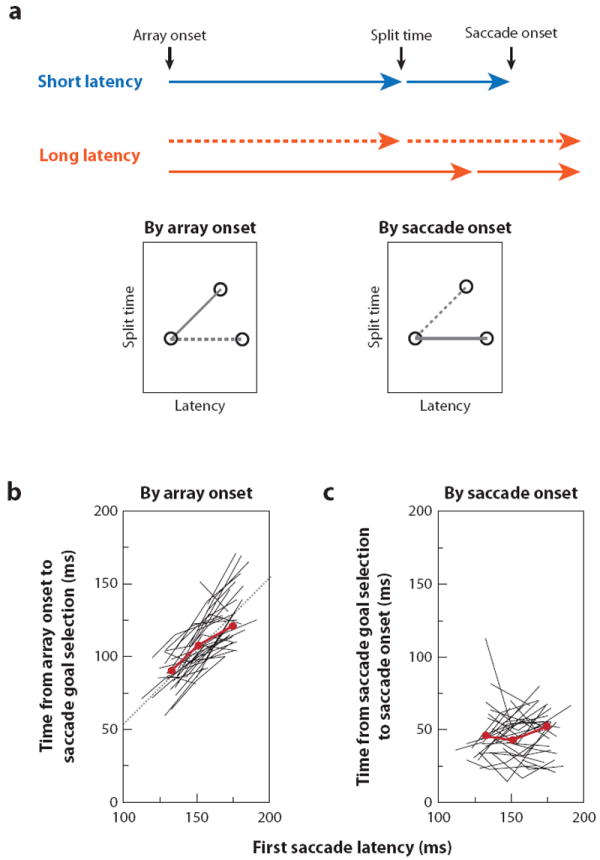
For many years there has been a debate about the role of the parietal lobe in the generation of behavior. Does it generate movement plans (intention) or choose objects in the environment for further processing? To answer this, we focus on the lateral intraparietal area (LIP), an area that has been shown to play independent roles in target selection for saccades and the generation of visual attention. Based on results from a variety of tasks, we propose that LIP acts as a priority map in which objects are represented by activity proportional to their behavioral priority. We present evidence to show that the priority map combines bottom-up inputs like a rapid visual response with an array of top-down signals like a saccade plan. The spatial location representing the peak of the map is used by the oculomotor system to target saccades and by the visual system to guide visual attention.


Conflict of interest statement

DISCLOSURE STATEMENT

The authors are not aware of any affiliations, memberships, funding, financial holdings, or any other conflicts of interests that might be perceived as affecting the objectivity of this review.

Figures

Figure 1
Figure 1
Posterior parietal cortex of human (left) and macaque monkey (right). (a) The human posterior parietal cortex (PPC) is divided by the intraparietal sulcus (IPS) into the superior parietal lobe (SPL) and the inferior parietal lobe (IPL). The IPL consists of the angular gyrus (Ang) and supramarginal gyrus (Smg) and borders the superior temporal gyrus (purple) at a region often referred to as the temporoparietal junction (TPJ). (b) The lunate and intraparietal sulci are opened up to show the locations of several extrastriate areas in addition to the visually responsive areas within the intraparietal sulcus. These include the parieto-occipital area (PO), the posterior intraparietal area (PIP), the medial intraparietal area (MIP), the lateral intraparietal area (LIP), the ventral intraparietal area (VIP), and the anterior intraparietal area (AIP). Adapted from Husain & Nachev (2007) and Colby et al. (1988).
Figure 2
Figure 2
Effect of a recent flash on the response of an LIP neuron in the stable array task. (a) In this task the eight objects remained on the screen for the duration of the experiment. The monkey initiated the trial by fixating a small spot (FP) positioned such that none of the stable objects appeared in the receptive field (RF). After a short delay the fixation point jumped to the center of the array, bringing one of the objects into the receptive field. (b) The response to a single stimulus flashing in the receptive field during a fixation task. No other stimuli were present on the screen. Activity is aligned on the stimulus onset. (c) The response to the same stimulus, as part of the stable array, moving into the receptive field by a saccade. Activity is aligned by the end of the saccade. (d) A saccade brings a recently flashed stimulus into the receptive field. The stimulus appears approximately 500 ms before the saccade, and the data are aligned by the end of the saccade. The gray bars beneath the spike density functions show when, during the trial, the stimulus was in the receptive field of the neuron. The up arrows represent the onset of a flashed stimulus, and the down arrows represent its disappearance. Activity is aligned on the saccade end. Adapted from Kusunoki et al. (2000).
Figure 3
Figure 3
Responses of a population of LIP neurons to salient events (perturbations) that occurred 200 ms before stimuli were revealed in a covert visual search task. Data show the responses to perturbations as the difference between responses on trials with and without the perturbation occurring on trials in which the animal knew that the target would appear on the SAME or OPPOSITE location as the perturbation. The perturbations were: an increase in luminance (INT+); a change in color (COL); a decrease in luminance (INT-); the appearance of frame surrounding one pattern (FRAME); and a back-and-forth radial movement (MOVE). Perturbation onset (PB.ON) is indicated by the solid vertical line; and search target onset (TG.ON) is indicated by the dashed vertical line. Reproduced from Balan & Gottlieb (2006) with permission.
Figure 4
Figure 4
Response of an LIP neuron in a stable array task requiring a saccade to a cued object. While the monkey was fixating outside of the array, a cue was flashed. The fixation point (FP) then jumped into the center of the array bringing an object into the receptive field. The animal then waited until the fixation point was extinguished and made a saccade to the cued object. (a) The cued object was within the receptive field after the first saccade; (b) the same object, this time not the target of the saccade, was brought into the receptive field by the first saccade. Each trio of raster plots shows the response of the neuron in the same trials synchronized on the cue (left), first saccade beginning (middle) and second saccade beginning (right). Adapted from Kusunoki et al. (2000) with permission.
Figure 5
Figure 5
LIP responses to a task-irrelevant popout distractor. (a) Responses of a single neuron to the appearance of an array object in the receptive field are plotted against time from target onset. Gray trace: response to a non-popout distractor in the receptive field when the monkey made a saccade to the target elsewhere. Green trace: response to the popout distractor in the receptive field when the monkey made a saccade to the target elsewhere. (b) The response of each cell from a 50-ms epoch, starting 40 ms after the latency, to the non-popout distractor is plotted against the response of the same cell to the popout distractor. (c) Cell by cell correlation of response difference with saccade suppression. The percentage of trials in which the first saccade went to the popout distractor for each cell is plotted against the difference in the number of spikes between the responses (80–130 ms) to the non-popout and popout distractors for the cell recorded in that session. Reproduced from Ipata et al. (2006b) with permission.
Figure 6
Figure 6
Summation in LIP. (a) A cartoon illustration of the test of summation. The cognitive signal (Cog) was in trials in which the target was in the receptive field. k is the constant that accounts for the different strength of the cognitive signal (see text for more details). The visual signal (Vis) was in all trials in which a stimulus appeared in the receptive field. The saccadic signal (Sac) was in trials in which the monkey made a saccade toward the receptive field. (b) Single cell responses from trials in which the saccade was made to the target inside the receptive field (orange trace) are compared to the calculated signal obtained by summing the three components obtained in different trial types (blue trace). (c) The mean activity in 20-ms time intervals from 80 ms to 240 ms after array onset measured in saccade-to-target-in-the-receptive-field trials (abscissa) against the activity in the same intervals calculated from the other three trial types (ordinate) for the same single neuron. Least-squares correlation line is shown with a dashed gray line. (d) Bar plot of the distribution of the slopes, in degrees, from the regression analyses for each cell. Adapted from Ipata et al. (2009) with permission.
Figure 7
Figure 7
The task and data collected to compare the locus of attention with activity in LIP. (a) The task was based on a memory-guided saccade task, with a task-irrelevant distractor. In this task, before the fixation point (FP) was extinguished four rings appeared. One of the rings had a gap on either the left or the right (the probe). The monkey had to identify the side of ring that the gap was on and indicate it either by making the planned memory-guided saccade when the fixation point was extinguished (GO) or by canceling the saccade and maintaining fixation until the end of the trial (NOGO). (b) Pooled behavioral and physiological data from a single animal. The thin traces in the top panel show the animal’s behavioral performance plotted as normalized threshold. Points that are significantly beneath the black dashed line indicate an attentional advantage (*). The thick traces in the lower panel show the mean spike density functions from a population of 23 neurons (the width of the trace shows the SEM). Blue traces show data from trials in which the probe was placed at the target site, and the distractor had flashed elsewhere. Red traces show data from trials in which the probe was placed at the distractor site and the target had flashed elsewhere. The thin gray trace shows the result of a running statistical test showing when the thick red and blue traces were indistinguishable (gray block). The black bar shows the onset and duration of the distractor. From Bisley & Goldberg (2003) as modified in Bisley & Goldberg (2006) with permission.
Figure 8
Figure 8
The normalized population responses from one monkey to the GO, NOGO and null probes in the contrast sensitivity task (Figure 7a). The color coding indicates the direction of saccade plan and whether the trial was a GO or NOGO trial, and the thickness of the line indicates whether a GO, NOGO, or null probe was in the receptive field. Adapted from Bisley & Goldberg (2006) with permission.
Figure 9
Figure 9
Relationship between LIP activity and first saccadic latency. (a) An example of a short latency trial is compared to two possible long latency trials showing the two extreme possibilities in how the extra time is added to latency. In the upper (dashed) example, the time from array onset until the split time is identical, so all the variability in latency time comes in to the process later than LIP. This would suggest LIP is not involved in saccadic selection. In the lower (solid) example, the extra latency time comes before the split time in LIP. This would suggest that LIP is driving the saccade. The two sets of hypothesized results (dashed and solid) are plotted in the small panels comparing the split time calculated by array onset and split time calculated by saccade onset. (b) The time from array onset to split time is plotted against the mean first saccadic latency for each group for each cell. The dotted line shows an example slope of 1. (c) The time from the split time to saccade onset is plotted against the mean first saccadic latency for each group for each cell. The flat lines suggest that a saccade is generated a set time after a peak is identified in LIP. For (b) and (c), the black lines connect points from the same cell and the solid red lines connect the population means. Adapted from Ipata et al. (2006a) with permission.

References

    1. Andersen RA, Asanuma C, Cowan WM. Callosal and prefrontal associational projecting cell populations in area 7a of the macaque monkey: a study using retrogradely transported fluorescent dyes. J Comp Neurol. 1985;232:443–55. - PubMed
    1. Andersen RA, Buneo CA. Intentional maps in posterior parietal cortex. Annu Rev Neurosci. 2002;25:189–220. - PubMed
    1. Baizer JS, Desimone R, Ungerleider LG. Comparison of subcortical connections of inferior temporal and posterior parietal cortex in monkeys. Visual Neurosci. 1993;10:59–72. - PubMed
    1. Baizer JS, Ungerleider LG, Desimone R. Organization of visual inputs to the inferior temporal and posterior parietal cortex in macaques. J Neurosci. 1991;11:168–90. - PMC - PubMed
    1. Balan PF, Gottlieb J. Integration of exogenous input into a dynamic salience map revealed by perturbing attention. J Neurosci. 2006;26:9239–49. - PMC - PubMed

Publication types

LinkOut - more resources