Review
. 2010 Jan;233(1):126-45.
doi: 10.1111/j.0105-2896.2009.00863.x.

The U1-snRNP complex: structural properties relating to autoimmune pathogenesis in rheumatic diseases

Affiliations
Review

The U1-snRNP complex: structural properties relating to autoimmune pathogenesis in rheumatic diseases

Nicole H Kattah et al. Immunol Rev. 2010 Jan.

Abstract

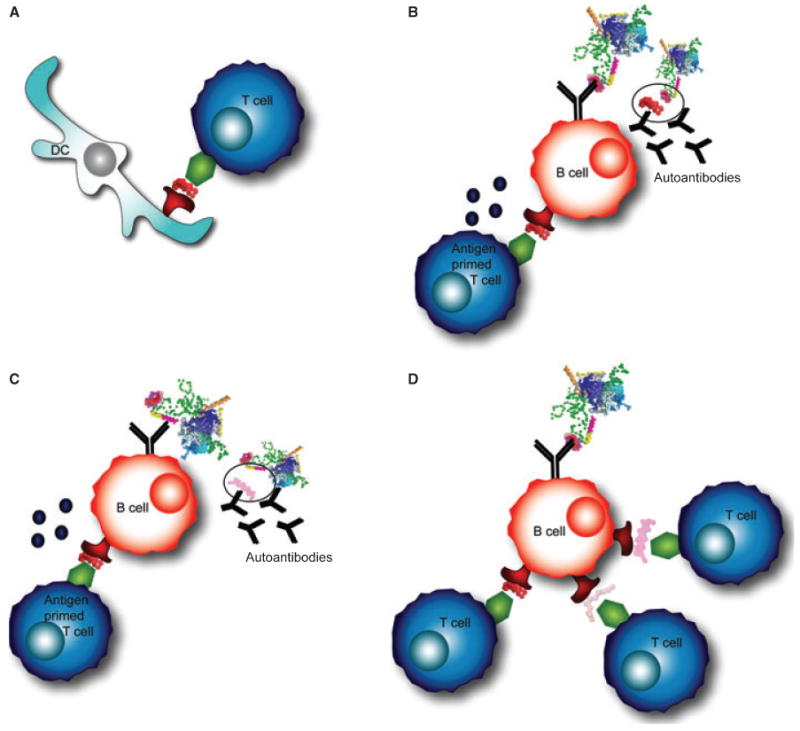
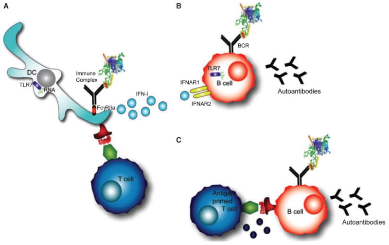
The U1 small nuclear ribonucleoprotein particle (snRNP) is a target of autoreactive B cells and T cells in several rheumatic diseases including systemic lupus erythematosus (SLE) and mixed connective tissue disease (MCTD). We propose that inherent structural properties of this autoantigen complex, including common RNA-binding motifs, B and T-cell epitopes, and a unique stimulatory RNA molecule, underlie its susceptibility as a target of the autoimmune response. Immune mechanisms that may contribute to overall U1-snRNP immunogenicity include epitope spreading through B and T-cell interactions, apoptosis-induced modifications, and toll-like receptor (TLR) activation through stimulation by U1-snRNA. We conclude that understanding the interactions between U1-snRNP and the immune system will provide insights into why certain patients develop anti-U1-snRNP autoimmunity, and more importantly how to effectively target therapies against this autoimmune response.


Figures

Fig. 1
Fig. 1. Immunoassays for detecting autoantibodies against U1-snRNP
(A) Hemagluttination, (B) The Immunodiffusion assay, (C) Immunofluorescence, (D) Immunoprecipitation, (E) Western blot, (F) ELISA, (G) Planar microarrays, (H) Bead-based arrays.
Fig. 2
Fig. 2. Modifications and autoimmune epitopes on U1-70K
(A) The U1-snRNP crystal structure (PDB ID: 3CW1) (7) was visualized using Pymol. In this depiction of the structure, U1-RNA is depicted in green, U1-70K in yellow, U1-C in orange, and the Sm proteins are displayed in blues. U1-A is not included in this depiction, as it was not part of the crystal complex in this structure. (B) The phosphorylation of serine residue 140 is a modification that occurs during apoptosis, and is shown in magenta. (C) The domain (100–180) of U1-70K is highlighted and is the RNA-binding domain of the protein. Notably the B-cell and T-cell epitope 131–151 is shown in red. The residues around this epitope are highlighted: 100–130 in magenta, and 152–180 in light pink. These regions contain additional B-cell and T-cell epitopes. (D) The domains of U1-70K include an N-terminal domain, RNA-binding domain, and serine/arginine repeats. During apoptosis, a 40 kDa fragment is cleaved at residue 341. B-cell and T-cell epitopes on U1-70K are shown.
Fig. 3
Fig. 3. Epitope spreading within the U1-snRNP complex
(A) Professional antigen-presenting cells, such as dendritic cells (DCs), prime antigen-specific T cells. DCs process the antigen complex and present peptides in MHC class II molecules on their surface. (B) The B-cell receptor (BCR) on a B cell recognizes the same complex, processes the antigen, and presents peptides to T cells. An antigen-primed T cell provides help to a cognate B cell, allowing the B cell to secrete antibodies specific for the same epitope as the BCR. (C) A B cell recognizing one portion of the antigen complex may present a peptide to T cells from another portion of the complex and receive T-cell help. This interaction results in autoantibody production to an epitope on the complex that is different from the T cell epitope. (D) A B cell that recognizes one portion of the complex could internalize, process, and present peptides to T cells that recognize epitopes from other portions of the U1-snRNP particle.
Fig. 4
Fig. 4. T-cell-independent and T-cell-dependent mechanisms of autoantibody production
(A) Dendritic cells internalize immune complexes through FcγRIIα. RNA contained in the immune complex stimulates TLR7, resulting in type-I interferon production (IFN-I). In addition, DCs present peptides from the processed antigen complex on their surface to T cells. (B) T-cell-independent B-cell activation occurs in B cells that are stimulated through the B-cell receptor (BCR) and TLR7 by RNA-containing antigens. In addition, interferons signal through IFNAR receptors on the B-cell surface. These signals can induce autoantibody production against the RNA-containing protein complex in some models. (C) T-cell-dependent B-cell activation occurs through the typical T-B cognate interaction that provides T-cell help and leads to autoantibody production.

Similar articles

Cited by

References

    1. von Muhlen CA, Tan EM. Autoantibodies in the diagnosis of systemic rheumatic diseases. Semin Arthritis Rheum. 1995;24:323–358. - PubMed
    1. Holman H, Deicher HR. The reaction of the lupus erythematosus (L.E.) cell factor with deoxyribonucleoprotein of the cell nucleus. J Clin Invest. 1959;38:2059–2072. - PMC - PubMed
    1. Tan EM, Kunkel HG. Characteristics of a soluble nuclear antigen precipitating with sera of patients with systemic lupus erythematosus. J Immunol. 1966;96:464–471. - PubMed
    1. Sharp GC, et al. Association of autoantibodies to different nuclear antigens with clinical patterns of rheumatic disease and responsiveness to therapy. J Clin Invest. 1971;50:350–359. - PMC - PubMed
    1. van Venrooij WJ, Zendman AJ, Pruijn GJ. Autoantibodies to citrullinated antigens in (early) rheumatoid arthritis. Autoimmun Rev. 2006;6:37–41. - PubMed

Publication types

MeSH terms