Review
. 2010 Jan;233(1):181-202.
doi: 10.1111/j.0105-2896.2009.00865.x.

From HLA-B27 to spondyloarthritis: a journey through the ER

Affiliations
Review

From HLA-B27 to spondyloarthritis: a journey through the ER

Robert A Colbert et al. Immunol Rev. 2010 Jan.

Abstract

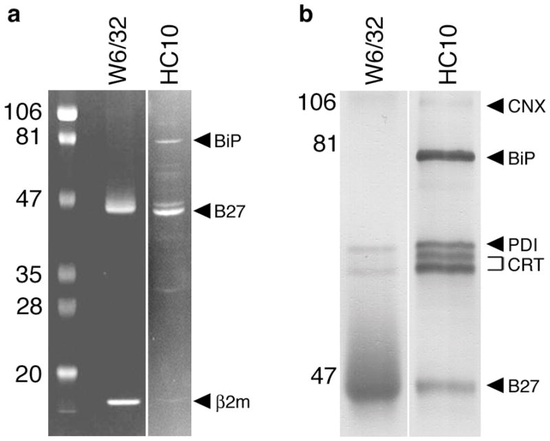
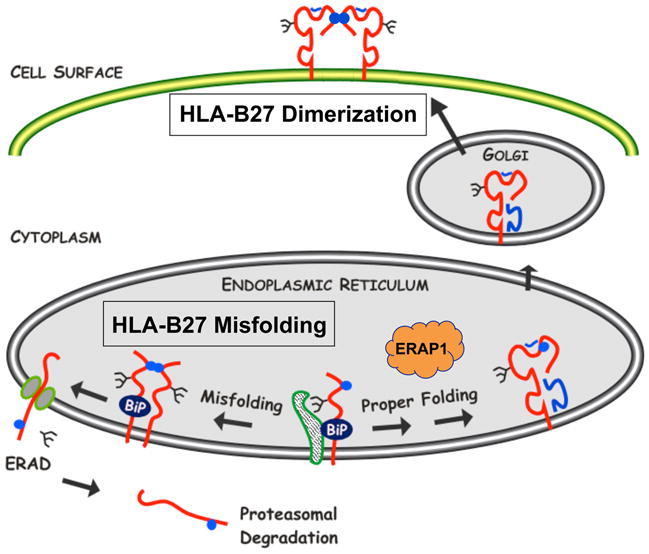
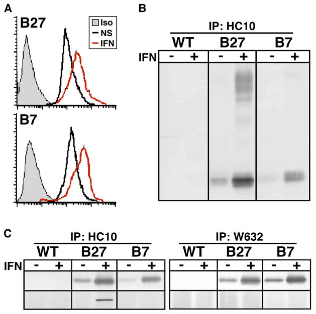
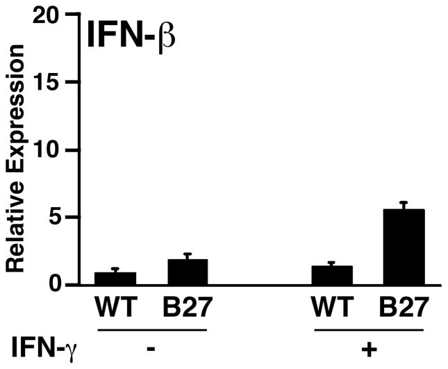
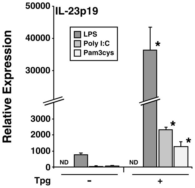
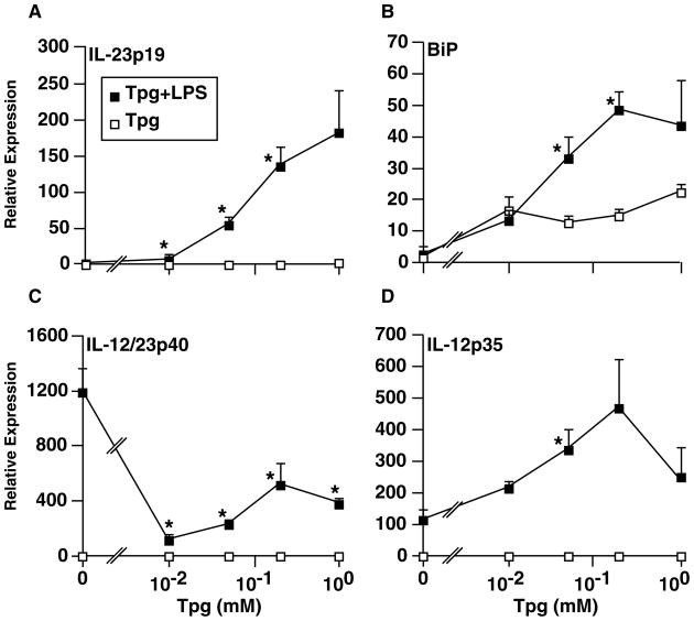
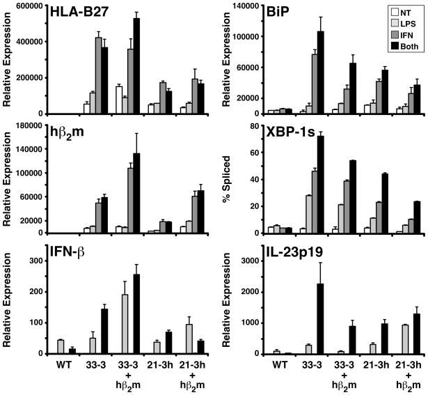
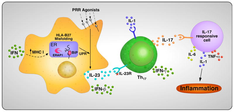
Almost four decades of research into the role of human leukocyte antigen-B27 (HLA-B27) in susceptibility to spondyloarthritis has yet to yield a convincing answer. New results from an HLA-B27 transgenic rat model now demonstrate quite convincingly that CD8(+) T cells are not required for the inflammatory phenotype. Discoveries that the HLA-B27 heavy chain has a tendency to misfold during the assembly of class I complexes in the endoplasmic reticulum (ER) and to form aberrant disulfide-linked dimers after transport to the cell surface have forced the generation of new ideas about its role in disease pathogenesis. In transgenic rats, HLA-B27 misfolding generates ER stress and leads to activation of the unfolded protein response, which dramatically enhances the production of interleukin-23 (IL-23) in response to pattern recognition receptor agonists. These findings have led to the discovery of striking T-helper 17 cell activation and expansion in this animal model, consistent with results emerging from humans with spondyloarthritis and the discovery of IL23R as an additional susceptibility gene for ankylosing spondylitis. Together, these results suggest a novel link between HLA-B27 and the T-helper 17 axis through the consequences of protein misfolding and open new avenues of investigation as well as identifying new targets for therapeutic intervention in this group of diseases.


Figures

Fig. 1
Fig. 1. ER chaperones associated with purified HLA-B27 heavy chains
HLA-B27 was purified from C1R.B27 cell lysates using sequential W6/32 and HC10 immunoaffinity columns. (A) Coomasie blue stain of HLA-B27 purified on W6/32 and HC10 columns separated on SDS-PAGE under reducing conditions. A reverse image is shown to enhance visualization of faint bands. HLA-B27, β2m, and BiP are indicated by arrows (right) and pre-stained markers (left) by their MW. BiP was identified by mass spectroscopic (MALDI-TOF) analysis of in-gel digested protein (data not shown). (B) Immunoblots of purified HLA-B27 performed using a cocktail of antibodies including AF-8 (anti-calnexin; CNX), anti-BiP (BiP), anti-PDI (PDI), anti-calreticulin (CRT), and 3B10.7 (anti-class I HC; B27). Prior to preparing the cocktail chaperones were first identified by immunoblotting with single antibodies (not shown). Calreticulin migrates as two forms indicated by the bracket. Band intensity in (A) reflects relative amount of protein stained with Coomassie blue.
Fig. 2
Fig. 2. HLA-B27 misfolding in the endoplasmic reticulum and dimers on the cell surface
Newly synthesized HLA-B27 heavy chains exhibit three features of protein misfolding: (i) prolonged and stable association with the ER chaperone BiP, (ii) formation of disulfide-linked dimers and possibly oligomers, and (iii) enhanced ERAD. ERAD involves dislocation of heavy chains from the lumen of the ER into the cytosol via the Sec61 complex and involves heavy chain deglycosylation and proteasomal degradation. HLA-B27 also has a tendency to form homodimers via endosomal recycling that can be expressed on the cell surface. Cell surface dimers shown here are in a folded conformation. Unfolded dimers as well as unfolded monomers also exist on the cell surface in varying amounts. Heavy chains are shown in red, with β2m and peptide in blue. Blue circles depict the unpaired Cys67 residue. Other Cys residues have also been implicated in aberrant disulfide bond formation. The figure is reproduced in a modified form from ref. (7) with permission.
Fig. 3
Fig. 3. Upregulation of MHC class I heavy chains with IFN-γ exacerbates HLA-B27 misfolding in rat macrophages
(A) Flow cytometric analysis of untreated (NS) and IFN-γ treated (IFN) (100 U/ml for 24 h) HLA-B27/hβ2m transgenic (B27) and HLA-B7/hβ2m transgenic (B7) rat macrophages stained with W6/32 or isotype (Iso) control antibody. (B and C) Immunoblots of HC10 (B) and HC10- and W6/32- (C) immunoprecipitated (IP) heavy chains from wildtype (WT), HLA-B27/hβ2m transgenic (B27), and HLA-B7/hβ2m transgenic (B7) macrophages treated without (−) and with (+) IFN-γ (IFN) for 24 h. (B) Non-reducing SDS-PAGE showing monomers (lower band) and disulfide-linked oligomers (upper bands) visualized with 3B10.7, a rat mAb that recognizes HLA class I heavy chains. (C) Reducing SDS-PAGE showing monomers (upper panel) visualized with 3B10.7, and co-precipitating BiP visualized with anti-KDEL (lower panel). W6/32 immunoprecipitates separated on non-reducing SDS-PAGE shows no evidence of disulfide-linked oligomers (unpublished observations). Some of the data in this figure were published previously in ref. (43) and are reprinted with permission.
Fig. 4
Fig. 4. IFN-β transcripts are upregulated in HLA-B27/hβ2m transgenic macrophages with UPR activation
BM macrophages derived from 4-week-old HLA-B27/hβ2m transgenic rats remained untreated (−) or were treated with 100 U/ml IFN-γ (+) for 24 h. RNA was isolated and IFN-β transcripts quantified with quantitative real time RT-PCR (qPCR). Results are normalized to actin mRNA. Bars represent mean of triplicate cultures with standard deviation shown. P < 0.05 for B27 vs. WT, and for B27 (+) vs. B27 (−), but P > 0.05 for WT (−) vs. WT (+). Microarray results from this experiment are shown in Fig. 7 in ref. (42).
Fig. 5
Fig. 5. Multiple TLR ligands synergistically upregulate IL-23p19 mRNA in macrophages experiencing ER stress
BM-derived macrophages from B6 mice remained untreated (−) or were pre-treated with thapsigargin (Tpg) (+) at a concentration of 1 μM for 1 h, then a TLR4 ligand (LPS, 10 ng/ml) was added for an additional 3 h, a TLR3 ligand (Poly I:C, 10 μg/ml) for 5 h, or a TLR2 ligand (Pam3cys, 1 μg/ml) for 3 h. RNA was isolated and IL-23p19 mRNA quantified with qPCR. All values have been normalized to β-actin mRNA. Bars represent mean of triplicate cultures with standard deviation shown. ND, not detected. Asterisks indicate P < 0.05 compared to respective (−) Tpg controls.
Fig. 6
Fig. 6. Effects of ER stress on IL-12/23 subunit mRNA induction by LPS
BM-derived macrophages from B6 mice were untreated or pre-treated with varying concentrations of thapsigargin (Tpg) (0 – 1 μM) for 1 hr, followed by LPS (10 ng/ml) for an additional 2 h. RNA was isolated and mRNA quantified with qPCR. All values have been normalized to GAPDH mRNA. Each data point is the mean of triplicate cultures with standard deviation shown. Effects of LPS alone are shown at ‘0’ Tpg. Asterisks indicate P < 0.05 compared to respective controls without LPS.
Fig. 7
Fig. 7. Additional hβ2m does not prevent UPR activation in HLA-B27/hβ2m-expressing rat macrophages
BM macrophages were derived from WT and 4 strains of HLA-B27/hβ2m transgenic rats that have different copy numbers of HLA-B27 and hβ2m transgenes. 33-3 rats have 55 copies of HLA-B27 and 66 copies of hβ2m; 33-3 × 283-2 (33-3 + hβ2m) have 35 additional copies of hβ2m from the 283-2 line. 21-3 rats have 20 copies of HLA-B27 and 15 copies of hβ2m; 21-3 × 283-2 (21-3 + hβ2m) have 35 additional copies of hβ2m. Macrophages remained untreated (NT) or were treated with 100 U/ml IFN-γ (IFN) for 24 h, 10 ng/ml LPS (LPS) for the last 3 h of culture, or IFN-γ + LPS (Both) with LPS present for the last 3 h of the 24 h IFN-γ treatment. RNA was isolated and mRNA for the various genes quantified with qPCR. Results are normalized to actin mRNA. XBP-1s is the percent of XBP1 mRNA transcripts that are spliced, and was measured as described previously (43, 65). Bars represent mean of triplicate cultures with standard deviation shown.
Fig. 8
Fig. 8. Hypothetical mechanism linking HLA-B27 misfolding and other disease associated genes to the IL-23/IL-17 axis
Cytokines that upregulate the MHC class I expression and assembly pathway, such as IFNs and/or TNF-α, may trigger an UPR in antigen-presenting cells of HLA-B27-expressing individuals when a critical threshold of misfolding is reached. ERAP1 polymorphisms could influence the efficiency of HLA-B27 folding, its propensity to misfold, and/or its peptide repertoire, through its effect on peptide trimming in the ER. The UPR will polarize cells responding to PRR agonists toward the production of greater amounts of IL-23 over IL-12, which in turn provides a stimulus for Th17 survival and activation in individuals with permissive IL23R polymorphisms. Th17 cells are shown producing IFN-γ, which has been documented in several situations, but would also be expected to come from Th1 cells. IL-1 locus polymorphisms (possibly IL1A) could play a role in Th17 development as well as in other inflammatory processes. This schema is meant as a framework to provide testable hypotheses regarding the role of various polymorphic susceptibility genes in the pathogenesis of spondyloarthritis. Additional susceptibility genes that fit within this schema are being identified (unpublished data). This figure is reproduced in a modified form from “Molecular Mechanisms of Spondyloarthropathies”, edited by Carlos López-Larrea and Roberto Diaz-Peña (c2009 Landes Bioscience and Springer Science+Business Media) with permission.

References

    1. Helmick CG, et al. Estimates of the prevalence of arthritis and other rheumatic conditions in the United States. Part I. Arthritis Rheum. 2008;58:15–25. - PubMed
    1. Braun J, Sieper J. Ankylosing spondylitis. Lancet. 2007;369:1379–1390. - PubMed
    1. Caffrey MF, James DC. Human lymphocyte antigen association in ankylosing spondylitis. Nature. 1973;242:121. - PubMed
    1. Brewerton DA, Hart FD, Nicholls A, Caffrey M, James DCO, Sturrock RD. Ankylosing spondylitis and HL-A 27. Lancet. 1973;1:904–907. - PubMed
    1. Schlosstein L, Terasaki PI, Bluestone R, Pearson CM. High association of an HL-A antigen, W27, with ankylosing spondylitis. N Engl J Med. 1973;288:704–706. - PubMed

Publication types

MeSH terms