Review
. 2010 Jan;233(1):233-55.
doi: 10.1111/j.0105-2896.2009.00859.x.

Fibroblast-like synoviocytes: key effector cells in rheumatoid arthritis

Affiliations
Review

Fibroblast-like synoviocytes: key effector cells in rheumatoid arthritis

Beatrix Bartok et al. Immunol Rev. 2010 Jan.

Abstract

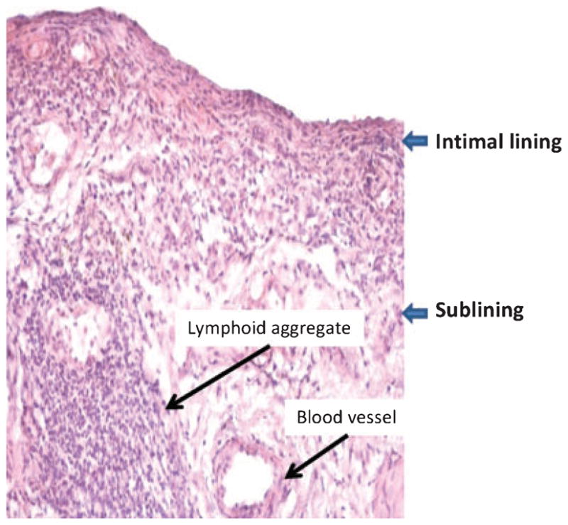
Rheumatoid arthritis (RA) remains a significant unmet medical need despite significant therapeutic advances. The pathogenesis of RA is complex and includes many cell types, including T cells, B cells, and macrophages. Fibroblast-like synoviocytes (FLS) in the synovial intimal lining also play a key role by producing cytokines that perpetuate inflammation and proteases that contribute to cartilage destruction. Rheumatoid FLS develop a unique aggressive phenotype that increases invasiveness into the extracellular matrix and further exacerbates joint damage. Recent advances in understanding the biology of FLS, including their regulation regulate innate immune responses and activation of intracellular signaling mechanisms that control their behavior, provide novel insights into disease mechanisms. New agents that target FLS could potentially complement the current therapies without major deleterious effect on adaptive immune responses.


Figures

Fig. 1
Fig. 1. Histopathologic appearance of rheumatoid arthritis(RA) synovial tissue
The synovium in RA is marked by intimal lining hyperplasia, infiltrating mononuclear cells in the sublining, and occasional lymphoid aggregates. From Reference (142).
Fig. 2
Fig. 2. p53 protein expression in synovial tissue
Immunohistochemistry was performed on synovial tissue from patients with (A) rheumatoid arthritis (RA) and (B) osteoarthritis (OA) to detect p53 protein. p53 was detected in the intimal lining and sublining infiltrating leukocytes. The p53 protein expression is significantly higher in the RA synovial tissue compared with OA synovium. From Reference (24).
Fig. 3
Fig. 3. Fibroblast-like synoviocytes (FLS) have intrinsic capacity to form a lining layer in vitro organ culture model
Cultured FLS were cultured in an artificial matrix and cultured for 3 weeks. The three-dimensional structure was fixed with 2% paraformaldehyde, embedded in paraffin and sections were stained with hematoxylin and eosin. FLS migrated to the surface of the matrix to establish a lining layer at the matrix-liquid interface (A). Immunohistochemistry was performed with cadherin-11 specific antibody and isotype control (B). Cadherin-11 specific staining was localized in the lining layer. From Reference (31).
Fig. 4
Fig. 4. Effect of p53 deficiency on fibroblast-like synoviocytes (FLS) aggressive phenotype
FLS derived from normal or rheumatoid synovial tissue were transduced with human papilloma virus type 18 (HPV-18) encoding E6 gene, which inactivates p53. The FLS were co-implanted with cartilage into SCID mice and harvested 60 days later. RA FLS (A) adhered to and invaded the matrix compared with normal FLS (C) which did not invade. Cartilage invasion was increased when RA FLS was transduced with HPV-18 encoding E6 gene (B). The normal FLS (C) acquired an RA-like invasive phenotype when p53 protein was inactivated with E6 (D). From Reference (42).
Fig. 5
Fig. 5. Apoptosis of RA FLS induced by PUMA overexpression
(A) RA FLS were transfected with pCEP4, hemagglutinin-tagged, full-length PUMA expression vector (HA-PUMA), or HA-tagged PUMA expression vector with a deletion of the Bcl-2 homology 3 domain (HA-PUMAdBH3). Data are presented as the percentage of nonviable cells. HA-PUMA-transfected FLS showed significantly more dead cells compared with pCEP4- or HA-PUM-AdBH3-transfected cells (*P < 0.01). (B) Comparison of apoptosis induced by pCEP4, HA-PUMA, or HA-PUMAdBH3 in RA and OA FLS lines. The extent of PUMA-induced apoptosis was the same in both cell lines. *P < 0.05 compared with controls. (C) RA FLS were transfected with pCEP4, HA-PUMA, or HA-PUMAdBH3 and DNA fragmentation, another measure of apoptosis, was measured by ELISA. Significant induction of DNA fragmentation was noted in HA-PUMA-transfected cells compared with pCEP4-transfected cells (*P < 0.05). (D) FLS transfected with pCEP4, HA-PUMA, or HA-PUMAdBH3 were cultured in chamber slides. The cells were then immunostained for activated caspase 3. HA-PUMA-transfected FLS showed significantly more activated caspase 3-positive cells compared with pCEP4- or HA-PUMAdBH3-transfected cells (P < 0.01). From reference (84).
Fig. 6
Fig. 6. TLR-3 mediated MMPs and type I IFN gene expression in fibroblast-like synoviocytes (FLS)
MMPs gene expression: After TLR-3 receptor stimulation by synthetic ligand poly (I:C), two independent pathways are activated, MKK7/JNK and IKKε/TBK1. Both of these complexes phosphorylate c-Jun, leading to increased AP-1 activation. JNK-mediated c-Jun activation and AP-1-mediated gene expression of MMPs require MKK7. IKKε, independent of JNK can also phosphorylate c-Jun after forming a complex with TBK1 and NAP1. Type I IFN gene expression: Transcriptional activation of type I IFN genes require formation of an enhanceosome, an interaction between transcription factors ATF2/c-Jun and IRF3. In FLS, in response to TLR-3 activation, IRF3 serves as a substrate for both JNK and IKKε, followed by phosphorylation, dimerization and translocation to the nucleus. JNK-mediated IRF3 activation and IFN gene expression requires MKK7. MKK4 in complex with JNK also contributes to type I IFN gene expression through ATF2 phosphorylation and formation of ATF2/c-Jun complex. TLR3, Toll-like receptor; MKK4, MAPK kinase 4; MKK7, MAPK kinase 7; JNK, c-Jun N-terminal kinase; TBK1, TANK-binding kinase; NAP1, NAK-associated protein-1; IKKe, IKK-related kinase; IRF3, interferon regulatory factor 3; c-JUN, component of AP-1 transcription factor; ATF2, activated transcription factor 2; MMPs, matrix metalloproteinases; Type I IFNs, interferon α, β and interferon-stimulated genes.

Similar articles

Cited by

References

    1. Gabriel SE, Michaud K. Epidemiological studies in incidence, prevalence, mortality, and comorbidity of the rheumatic diseases. Arthritis Res Ther. 2009;11:229. - PMC - PubMed
    1. Firestein GS. Evolving concepts of rheumatoid arthritis. Nature. 2003;423:356–361. - PubMed
    1. Bresnihan B, Flanagan AM. Synovium. In: Firestein GS, Budd RC, Harris T, McInnes IB, Ruddy S, Sergent JS, editors. Kelly’s Textbook of Rheumatology. 8. Philadelphia, PA: Saunders Elsevier; 2009. pp. 23–37.
    1. Edwards JC, Willoughby DA. Demonstration of bone marrow derived cells in synovial lining by means of giant intracellular granules as genetic markers. Ann Rheum Dis. 1982;41:177–182. - PMC - PubMed
    1. Firestein GS. Invasive fibroblast-like synoviocytes in rheumatoid arthritis. Passive responders or transformed aggressors? Arthritis Rheum. 1996;39:1781–1790. - PubMed