Review
. 2010 Jan;233(1):286-300.
doi: 10.1111/j.0105-2896.2009.00849.x.

Transcriptional regulation of bone and joint remodeling by NFAT

Affiliations
Review

Transcriptional regulation of bone and joint remodeling by NFAT

Despina Sitara et al. Immunol Rev. 2010 Jan.

Abstract

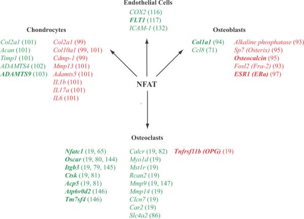
Osteoporosis and arthritis are highly prevalent diseases and a significant cause of morbidity and mortality worldwide. These diseases result from aberrant tissue remodeling leading to weak, fracture-prone bones or painful, dysfunctional joints. The nuclear factor of activated T cells (NFAT) transcription factor family controls diverse biologic processes in vertebrates. Here, we review the scientific evidence that links NFAT-regulated gene transcription to bone and joint pathology. A particular emphasis is placed on the role of NFATs in bone resorption and formation by osteoclasts and osteoblasts, respectively. In addition, emerging data that connect NFATs with cartilage biology, angiogenesis, nociception, and neurogenic inflammation are explored. The goal of this article is to highlight the importance of tissue remodeling in musculoskeletal disease and situate NFAT-driven cellular responses within this context to inspire future research endeavors.


Figures

Fig. 1
Fig. 1. Regulation of gene expression by NFAT in endothelial cells, chondrocytes, osteoblasts, and osteoclasts
Genes included on this list were shown to be NFAT regulated by promoter reporter assays, ChIP experiments, overexpression studies, or differential regulation in wildtype versus NFAT-deficient cells. Genes whose expression is augmented by NFAT are shown in green. Genes whose expression is repressed by NFAT are shown in red. The promoters of the genes in bold have been shown by ChIP to be direct NFAT targets.
Fig. 2
Fig. 2. Mutant SH3BP2 promotes osteoclastogenesis via NFATc1
(A) Micro-computed tomography (μ-CT) images of the distal femoral metaphysis of wildtype (Sh3bp2+/+) and cherubism mice (Sh3bp2KI/KI) either sufficient (Nfatc1+/+) or deficient (NFATc1Δ/Δ) in NFATc1. Note the dramatic osteoporosis in Sh3bp2KI/KI mice compared with wildtype mice (upper right and upper left panels). In the absence of NFATc1 (lower right panel), osteoporosis is reversed in Sh3bp2KI/KI mice and these mice display osteopetrosis similar to Sh3bp2+/+ lacking NFATc1 (lower left panel). (B) Signals emanating from RANK during osteoclast differentiation induce the expression and activation of NFATc1, which promotes additional NFATc1 expression by autoregulation of the Nfatc1 promoter (65). (C) Mutant SH3BP2 (P416R) augments the osteoclast differentiation program by driving NFATc1 autoamplification (19). Image in (A) courtesy of Dr. Ted Gross from the University of Washington.
Fig. 3
Fig. 3. SLC4A2/AE2 is necessary for bone resorption by osteoclasts
(A) Diagram of the osteoclast acidification pathway. Carbonic anhydrase II (CAII) generates carbonic acid (H2CO3), which dissociates into a bicarbonate ion (HCO3) and a proton (H+). The proton is secreted into the resorption lacuna via a vacuolar-type H+ATPase located in the ruffled border of the apical membrane. A chloride (Cl) equivalent is also released via CLCN7 into the resorption lacuna to maintain electroneutrality. Excess cytoplasmic HCO3 is exchanged for Cl via SLC4A2/AE2 located in the basolateral membrane, which prevents intracellular alkalinization (85, 86). (B) μCT reconstruction of femurs from a wild-type mouse (left) and a mouse deficient in the HCO3/Cl exchanger, SLC4A2/AE2 (right). Absence of AE2 perturbs the bone resorbing function of osteoclasts, leading to a massive accumulation of trabecular bone (pseudocolored in orange). Image in (B) courtesy of Nicholas James Brady from the μCT facility at the Harvard School of Dental Medicine.

Similar articles

Cited by

References

    1. Harris ED., Jr The bone and joint decade: a catalyst for progress. Arthritis Rheum. 2001;44:1969–1970. - PubMed
    1. Woolf AD, Pfleger B. Burden of major musculoskeletal conditions. Bull World Health Organ. 2003;81:646–656. - PMC - PubMed
    1. Karsenty G, Wagner EF. Reaching a genetic and molecular understanding of skeletal development. Dev Cell. 2002;2:389–406. - PubMed
    1. Boyce BF, Xing L. Biology of RANK, RANKL, and osteoprotegerin. Arthritis Res Ther. 2007;9(Suppl):S1. - PMC - PubMed
    1. Phillips K, Aliprantis A, Coblyn J. Strategies for the prevention and treatment of osteoporosis in patients with rheumatoid arthritis. Drugs Aging. 2006;23:773–779. - PubMed

Publication types

MeSH terms