

Langerhans cells and more: langerin-expressing dendritic cell subsets in the skin
- PMID: 20193016
- PMCID: PMC2907488
- DOI: 10.1111/j.0105-2896.2009.00886.x
Langerhans cells and more: langerin-expressing dendritic cell subsets in the skin
Abstract
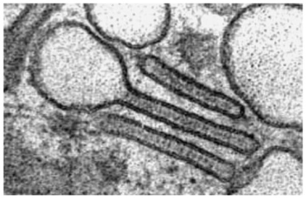
Langerhans cells (LCs) are antigen-presenting dendritic cells (DCs) that reside in epithelia. The best studied example is the LC of the epidermis. By electron microscopy, their identifying feature is the unique rod- or tennis racket-shaped Birbeck granule. The phenotypic hallmark is their expression of the C-type lectin receptor langerin/CD207. Langerin, however, is also expressed on a recently discovered population of DC in the dermis and other tissues of the body. These 'dermal langerin(+) dendritic cells' are unrelated to LCs. The complex field of langerin-negative dermal DCs is not dealt with here. In this article, we briefly review the history, ontogeny, and homeostasis of LCs. More emphasis is laid on the discussion of functional properties in vivo. Novel models using genetically engineered mice are contributing tremendously to our understanding of the role of LCs in eliciting adaptive immune responses against pathogens or tumors and in inducing and maintaining tolerance against self antigens and innocuous substances in vivo. Also, innate effector functions are increasingly being recognized. Current activities in this area are reviewed, and possibilities for future exploitation of LC in medicine, e.g. for the improvement of vaccines, are contemplated.
Figures

References
-
- Steinman RM. Dendritic cells: versatile controllers of the immune system. Nat Med. 2007;13:1155–1159. - PubMed
-
- Steinman RM, Banchereau J. Taking dendritic cells into medicine. Nature. 2007;449:419–426. - PubMed
-
- Steinman RM, Hemmi H. Dendritic cells: translating innate to adaptive immunity. Curr Top Microbiol Immunol. 2006;311:17–58. - PubMed
-
- Langerhans P. Über die Nerven der menschlichen Haut. Virchows Arch [A] 1868;44:325–337.
Publication types
MeSH terms
Substances
Grants and funding
LinkOut - more resources
Full Text Sources
Other Literature Sources
