Review
. 2010 Mar;234(1):142-62.
doi: 10.1111/j.0105-2896.2009.00881.x.

Unraveling the functions of plasmacytoid dendritic cells during viral infections, autoimmunity, and tolerance

Affiliations
Review

Unraveling the functions of plasmacytoid dendritic cells during viral infections, autoimmunity, and tolerance

Melissa Swiecki et al. Immunol Rev. 2010 Mar.

Abstract

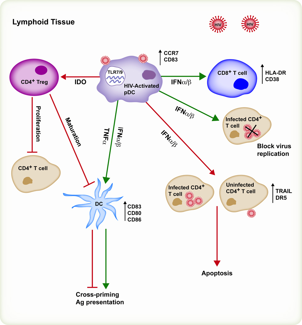
Plasmacytoid dendritic cells (pDCs) are bone marrow-derived cells that secrete large amounts of type I interferon (IFN) in response to viruses. Type I IFNs are pleiotropic cytokines with antiviral activity that also enhance innate and adaptive immune responses. Viruses trigger activation of pDCs and type I IFN responses mainly through the Toll-like receptor pathway. However, a variety of activating and inhibitory pDC receptors fine tune the amplitude of type I IFN responses. Chronic activation and secretion of type I IFN in the absence of infection can promote autoimmune diseases. Furthermore, while activated pDCs promote immunity and autoimmunity, resting or alternatively activated pDCs may be tolerogenic. The various roles of pDCs have been extensively studied in vitro and in vivo with depleting antibodies. However, depleting antibodies cross-react with other cell types that are critical for eliciting protective immunity, potentially yielding ambiguous phenotypes. Here we discuss new approaches to assess pDC functions in vivo and provide preliminary data on their potential roles during viral infections. Such approaches would also prove useful in the more specific evaluation of how pDCs mediate tolerance and autoimmunity. Finally, we discuss the emergent role of pDCs and one of their receptors, tetherin, in human immunodeficiency virus pathogenesis.


Figures

Fig. 1
Fig. 1. DT administration systemically depletes pDCs in BDCA-2-DTR mice
(A) Spleen, blood, lymph node (LN), and liver were analyzed for the expression of B220 and Siglec-H by flow cytometry 24 h after i.p. injection of PBS or DT. (B) pDCs-depleted mice have reduced type I IFN responses to CpG. BDCA-2-DTR mice were injected with CpG 2216 and DOTAP i.v. 24 h after i.p. injection of PBS or DT. Serum was collected 6 h later, and IFN-α levels were measured by ELISA.
Fig. 2
Fig. 2. Innate and adaptive responses to VSV-OVA in pDC-depleted mice
Mice were injected with PBS or DT on days −1 and 3 and infected with VSV-OVA (5×10⁶ pfu, i.v.). (A) Total cells per spleen on day 7 post-infection in control and pDC-depleted mice. (B) pDCs-depleted mice have a reduction in OVA-specific CD8+ T cells. Splenocytes from infected mice were stained with H-2Kb/OVA257–264 peptide tetramers and anti-CD8 and the frequencies and total numbers of antigen-specific CD8+ T cells were determined by flow cytometry. (C) Control and pDC-depleted mice produce similar amounts of systemic type I IFN and IL-12p40 following challenge with VSV-OVA. Serum was collected at various timepoints post-infection, and IFN-α and IL-12p40 levels were measured by ELISA and cytometric bead array, respectively. (D and E) pDC depletion does not affect DC maturation or antigen presentation in VSV-OVA-infected mice. (D) The expression of molecules on CD11chi DCs from spleens of naive mice or mice infected for 24 h with VSV-OVA were assessed by flow cytometry using antibodies to CD11c, CD80, CD86, MHC II, and CD40 (red histograms, DT; blue histograms, PBS; and black histograms, naïve mice). (E) CD11c+ cells from spleens of VSV-OVA-infected mice were enriched with MACS beads 24 h post-infection. CD11c+ cells were stained with anti-CD11c, -B220, -Siglec-H, and -CD8α antibodies and sorted into 3 populations: CD11c+CD8+ (CD8α+ DC), CD11c+CD8 (CD8α DCs), and Siglec-H+B220+ (pDCs). DC subsets were co-cultured at different ratios for 72 h with CD8+ T cells purified from OT-1 transgenic mice by negative selection. IFN-γ levels in culture supernatants were determined by cytometric bead array. (F) pDC-depleted mice infected with VSV-OVA produce less MIP-1β (CCL4) compared to control mice. Serum levels of MIP-1β were calculated by cytometric bead array. (G and H) Antibody responses to VSV-OVA in pDC-depleted mice. (G) Serum IL-6 levels were determined at 24 and 72 h post-infection by cytometric bead array. (H) Top panel: levels of OVA-specific IgM in serum on days 3 and 7 post-infection were assessed by ELISA. Bottom panel: Serum was collected on day 7 and used in standard virus neutralization assays with Vero cell monolayers. Briefly, VSV-OVA was incubated for 90 min with heat-inactivated, diluted mouse serum then added to Vero cells for 1 h. Wells were overlaid with methyl cellulose and plates were incubated for an additional 24 h. Cells were fixed with Carnoy’s fixative and stained with crystal violet. Virus incubated with medium or serum from naïve mice served as controls. (I) Virus titers in the brains of control or pDC-depleted mice on day 7. Confluent Vero cell monolayers were infected with brain homogenates for 1 h then overlaid with 1% methyl cellulose in complete medium. The overlay was flicked off 24 h later and cells were fixed with Carnoy’s fixative and stained with crystal violet. Data are combined from 2–3 separate experiments. Results depicted in bar graphs are the means +/− SEM. Student’s t test was used to calculate p values.
Fig. 3
Fig. 3. Innate and adaptive responses to LCMV in pDC-depleted mice
Mice were injected with PBS or DT on days −1 and 3 and infected i.p. with either 2×10⁵ or 1×10⁶ pfu of LCMV-Armstrong. (A) Control and pDC-depleted mice produce comparable levels of IFN-α, MCP-1, IL-6, and TNF-α during LCMV infection. Serum was collected at 24 and 72 h post-infection and analyzed by ELISA or cytometric bead array. (B) pDCs are not required for robust anti-LCMV CTL responses. Splenocytes were isolated on days 10 or 11 post-infection and stained with H-2Db/LCMVgp33–41 tetramers and anti-CD8 antibody, then analyzed by flow cytometry. The frequencies (top panels) and total numbers (middle panels) of antigen-specific T cells are shown. Splenocytes from day 10 infected mice (2×10⁵ pfu) were incubated in vitro for 6 h with LCMV peptides gp33–41 or gp276–286 and Brefeldin A (BFA). Cells were stained with antibodies to CD4, CD8, and IFN-γ and analyzed by flow cytometry. The frequencies and total numbers of cells producing IFN-γ are shown in the lower panels. Antigen-specific cytotoxicity was assessed using splenocytes from day 10 infected mice as effector cells and EL-4 cells pulsed or not pulsed with LCMV peptides gp33–41 or gp276–286 as targets in a standard 4-h 51Cr release assay. Results depicted in bar graphs are the means +/− SEM (n = 3 mice per dose/group).
Fig. 4
Fig. 4. Potential role of pDCs in the induction of antiviral CTL responses
pDCs sense viral nucleic acids via TLR7/9 in endosomal compartments. Upon activation, pDCs produce type I IFNs, IL-12, and chemokines such as MIP-1α and MIP-1β that attract naive CD8+ T cells to T-cell areas of secondary lymphoid tissues where they become virus-specific CTLs. Virus-specific CTLs proliferate and migrate to the periphery where they produce IFN-γ and kill virus-infected cells via perforin (Per) and granzyme (Gr). In our experimental settings, pDCs promote CD8+ T-cell responses mostly by secreting chemoattractants. However, pDCs may help CD8+ T-cell responses by secreting type I IFN and IL-12, which may act directly on T cells, or indirectly, by inducing the maturation of DCs.
Fig. 5
Fig. 5. The emergent roles of pDCs during HIV infection
It has been proposed that HIV-activated pDCs have beneficial and detrimental effects on HIV pathogenesis. Beneficial effects (green lines): pDCs secretion of type I IFNs and TNF-α promote the bystander maturation of DC which cross-prime and present antigens to T cells. Type I IFNs also limit viral replication in CD4+ T cells. Detrimental effects (red lines): HIV-activated pDCs express IDO, which induces Tregs that suppress CD4+ T-cell proliferation and DC maturation. Chronic pDC activation and type I IFN secretion has also been associated with the upregulation of activation markers on CD8+ T cells and the progressive depletion of infected and uninfected CD4+ T cells through apoptotic mechanisms such as TRAIL/DR5 or Fas/Fas ligand.
Fig. 6
Fig. 6. pDC depletion does not affect the replication of Listeria monocytogenes (LM) in organs of infected mice
BDCA-2-DTR mice were injected with PBS or DT on days −1 and 2 post-infection and infected with 1×10⁵ cfu of the EGD strain of LM i.p. Spleens and livers were collected on day 4 post-infection, homogenized and plated on BHI agar. Homogenates were serially diluted and plated in duplicate. Colony-forming units per organ were determined 24 h later. Bar graphs show the means +/− SEM (4 mice/group).

Similar articles

Cited by

References

    1. Colonna M, Trinchieri G, Liu YJ. Plasmacytoid dendritic cells in immunity. Nat Immunol. 2004;5:1219–1226. - PubMed
    1. Liu YJ. IPC: professional type 1 interferon-producing cells and plasmacytoid dendritic cell precursors. Annu Rev Immunol. 2005;23:275–306. - PubMed
    1. Muller-Hermelink HK, Stein H, Steinmann G, Lennert K. Malignant lymphoma of plasmacytoid T-cells. Morphologic and immunologic studies characterizing a special type of T-cell. Am J Surg Pathol. 1983;7:849–862. - PubMed
    1. Lennert K, Remmele W. [Karyometric research on lymph node cells in man. I. Germinoblasts, lymphoblasts & lymphocytes.] Acta Haematol. 1958;19:99–113. - PubMed
    1. Facchetti F, de Wolf-Peeters C, Mason DY, Pulford K, van den Oord JJ, Desmet VJ. Plasmacytoid T cells. Immunohistochemical evidence for their monocyte/macrophage origin. Am J Pathol. 1988;133:15–21. - PMC - PubMed

Publication types

MeSH terms