

Review
doi: 10.1111/j.0105-2896.2009.00880.x.
Microbe-dendritic cell dialog controls regulatory T-cell fate
Affiliations
- PMID: 20193027
- PMCID: PMC3404740
- DOI: 10.1111/j.0105-2896.2009.00880.x
Item in Clipboard
Review
Microbe-dendritic cell dialog controls regulatory T-cell fate
Immunol Rev.
2010 Mar.
Abstract
Each microenvironment is controlled by a specific set of regulatory elements that have to be finely and constantly tuned to maintain local homeostasis. These environments could be site specific, such as the gut environment, or induced by chronic exposure to microbes. Various populations of dendritic cells are central to the orchestration of this control. In this review, we discuss some new findings associating dendritic cells from defined compartments with the induction and control of regulatory T cells in the context of exposure to both commensal and pathogenic microbes.
Figures

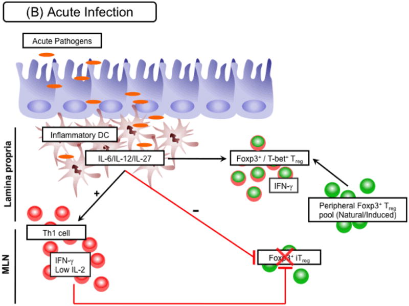
(A) Steady state/chronic infection. At steady state, several subsets of small intestine DCs are exposed to luminal antigens derived from gut flora or food. Of particular importance, CD103+ DCs metabolize vitamin A to retinoic acid (RA), which is able to favor conversion of Foxp3+ Tregs by directly acting on naive T cells and limiting effector cytokine release by activated T cells. Expansion of neoconverted Foxp3+ Tregs, along with other induced Foxp3+ Treg (iTregs) and natural Foxp3+ Tregs that make up the peripheral Treg pool, is further supported by the availability of IL-2 or other common γ chain cytokines (e.g. IL-7, IL-15). Subsequently, this compartment acts as one layer of regulation within the gut environment to limit local immune activation stimulated by luminal antigens. A similar scenario may occur in the context of chronic infection in which conversion needs to be maintained in the face of an ongoing immune response. As a consequence chronic pathogens may have co-evolved to release factors that enhance the conversion process, such as TGF-β. (B) Acute infection. In the presence of an acute infection, such as infection with Toxoplasma gondii, peripheral Foxp3+ Treg conversion is inhibited. This inhibition is in part mediated by the influx of inflammatory DC subsets into tissue sites. By releasing pro-inflammatory cytokines, including IL-12 or IL-27, these DCs support the expansion of IFN-γ secreting Th1 cells. IFN-γ directly inhibits the Foxp3+ Treg conversion process. In addition limitation of IL-2 in Th1 polarized environments hinders the development of this compartment. Moreover, IL-12 from inflammatory DC subsets can drive expression of T-bet within previously Foxp3+ Treg allowing them to adapt to the inflammatory within the tissue. All these mechanisms act together to favor a controlled effector response towards the invading pathogen. However, in highly inflammatory circumstances expression of T-bet may eventually drive IFN-γ production in regulatory T cells. In this situation the breakdown of the conversion process along with expression of effector cytokines by Foxp3+ Tregs may potentially result in severe pathogenesis.
(A) Steady state/chronic infection. At steady state, several subsets of small intestine DCs are exposed to luminal antigens derived from gut flora or food. Of particular importance, CD103+ DCs metabolize vitamin A to retinoic acid (RA), which is able to favor conversion of Foxp3+ Tregs by directly acting on naive T cells and limiting effector cytokine release by activated T cells. Expansion of neoconverted Foxp3+ Tregs, along with other induced Foxp3+ Treg (iTregs) and natural Foxp3+ Tregs that make up the peripheral Treg pool, is further supported by the availability of IL-2 or other common γ chain cytokines (e.g. IL-7, IL-15). Subsequently, this compartment acts as one layer of regulation within the gut environment to limit local immune activation stimulated by luminal antigens. A similar scenario may occur in the context of chronic infection in which conversion needs to be maintained in the face of an ongoing immune response. As a consequence chronic pathogens may have co-evolved to release factors that enhance the conversion process, such as TGF-β. (B) Acute infection. In the presence of an acute infection, such as infection with Toxoplasma gondii, peripheral Foxp3+ Treg conversion is inhibited. This inhibition is in part mediated by the influx of inflammatory DC subsets into tissue sites. By releasing pro-inflammatory cytokines, including IL-12 or IL-27, these DCs support the expansion of IFN-γ secreting Th1 cells. IFN-γ directly inhibits the Foxp3+ Treg conversion process. In addition limitation of IL-2 in Th1 polarized environments hinders the development of this compartment. Moreover, IL-12 from inflammatory DC subsets can drive expression of T-bet within previously Foxp3+ Treg allowing them to adapt to the inflammatory within the tissue. All these mechanisms act together to favor a controlled effector response towards the invading pathogen. However, in highly inflammatory circumstances expression of T-bet may eventually drive IFN-γ production in regulatory T cells. In this situation the breakdown of the conversion process along with expression of effector cytokines by Foxp3+ Tregs may potentially result in severe pathogenesis.
Similar articles
-
Tuning microenvironments: induction of regulatory T cells by dendritic cells.Immunity. 2008 Sep 19;29(3):362-71. doi: 10.1016/j.immuni.2008.08.005. Immunity. 2008. PMID: 18799144 Free PMC article. Review.
-
Regulation of intestinal homeostasis by dendritic cells.Immunol Rev. 2010 Mar;234(1):247-58. doi: 10.1111/j.0105-2896.2009.00872.x. Immunol Rev. 2010. PMID: 20193023 Review.
-
T-cell selection and intestinal homeostasis.Immunol Rev. 2014 May;259(1):60-74. doi: 10.1111/imr.12171. Immunol Rev. 2014. PMID: 24712459 Free PMC article. Review.
-
Regulatory T cells in the control of host-microorganism interactions (*).Annu Rev Immunol. 2009;27:551-89. doi: 10.1146/annurev.immunol.021908.132723. Annu Rev Immunol. 2009. PMID: 19302048 Review.
-
Eosinophils promote generation and maintenance of immunoglobulin-A-expressing plasma cells and contribute to gut immune homeostasis.Immunity. 2014 Apr 17;40(4):582-93. doi: 10.1016/j.immuni.2014.02.014. Immunity. 2014. PMID: 24745334
Cited by
-
Tregs and infections: on the potential value of modifying their function.J Leukoc Biol. 2011 Dec;90(6):1079-87. doi: 10.1189/jlb.0611271. Epub 2011 Sep 13. J Leukoc Biol. 2011. PMID: 21914856 Free PMC article. Review.
-
MyD88 signaling inhibits protective immunity to the gastrointestinal helminth parasite Heligmosomoides polygyrus.J Immunol. 2014 Sep 15;193(6):2984-93. doi: 10.4049/jimmunol.1401056. Epub 2014 Aug 11. J Immunol. 2014. PMID: 25114104 Free PMC article.
-
Role of Dendritic Cells in the Induction of Lymphocyte Tolerance.Front Immunol. 2015 Oct 20;6:535. doi: 10.3389/fimmu.2015.00535. eCollection 2015. Front Immunol. 2015. PMID: 26539197 Free PMC article. Review.
-
Liver cystic echinococcosis and human host immune and autoimmune follow-up: A review.World J Hepatol. 2017 Oct 28;9(30):1176-1189. doi: 10.4254/wjh.v9.i30.1176. World J Hepatol. 2017. PMID: 29109850 Free PMC article. Review.
-
Circulatory antigen processing by mucosal dendritic cells controls CD8(+) T cell activation.Immunity. 2013 Jan 24;38(1):153-65. doi: 10.1016/j.immuni.2012.09.018. Epub 2012 Dec 13. Immunity. 2013. PMID: 23246312 Free PMC article.
References
Publication types
MeSH terms
Substances
Grants and funding
LinkOut - more resources
Full Text Sources
Other Literature Sources
