. 2010 Mar 1:9:64.
doi: 10.1186/1475-2875-9-64.

Dual effect of Plasmodium-infected erythrocytes on dendritic cell maturation

Affiliations

Dual effect of Plasmodium-infected erythrocytes on dendritic cell maturation

Esther Bettiol et al. Malar J. .

Abstract

Background: Infection with Plasmodium is the cause of malaria, a disease characterized by a high inflammatory response in the blood. Dendritic cells (DC) participate in both adaptive and innate immune responses, influencing the generation of inflammatory responses. DC can be activated through different receptors, which recognize specific molecules in microbes and induce the maturation of DC.

Methods: Using Plasmodium yoelii, a rodent malaria model, the effect of Plasmodium-infected erythrocytes on DC maturation and TLR responses have been analysed.

Results: It was found that intact erythrocytes infected with P. yoelii do not induce maturation of DC unless they are lysed, suggesting that accessibility of parasite inflammatory molecules to their receptors is a key issue in the activation of DC by P. yoelii. This activation is independent of MyD88. It was also observed that pre-incubation of DC with intact P. yoelii-infected erythrocytes inhibits the maturation response of DC to other TLR stimuli. The inhibition of maturation of DC is reversible, parasite-specific and increases with the stage of parasite development, with complete inhibition induced by schizonts (mature infected erythrocytes). Plasmodium yoelii-infected erythrocytes induce a broad inhibitory effect rendering DC non-responsive to ligands for TLR2, TLR3, TLR4, TLR5, TLR7 and TLR9.

Conclusions: Despite the presence of inflammatory molecules within Plasmodium-infected erythrocytes, which are probably responsible for DC maturation induced by lysates, intact Plasmodium-infected erythrocytes induce a general inhibition of TLR responsiveness in DC. The observed effect on DC could play an important role in the pathology and suboptimal immune response observed during the disease. These results help to explain why immune functions are altered during malaria, and provide a system for the identification of a parasite-derived broad inhibitor of TLR-mediated signaling pathways.


Figures

Figure 1
Figure 1
P. yoelii lysates, but not whole P. yoelii-infected erythrocytes, induce DC maturation in vitro. (A) Bone marrow-derived DC were incubated with whole RBC (red), whole P. yoelii-iRBC (black), lysates of RBC (orange), or lysates of iRBC (purple).Ratio of red blood cell (RBC):DC was 30:1, an amount of lysate equivalent to a 30:1 ratio of whole parasites/DC was added to DC. Surface expression of maturation-associated receptors and MHC was determined by FACS after 24 h or 48 h. (B) DC derived from wt (circles) or MyD88-deficient (squares) mice were incubated with lysates of RBC (black symbols) or P. yoelii-iRBC (white symbols) at the indicated ratios of RBC:DC. The ratio indicates the amount of RBC or iRBC from which the lysate was derived. Expression of CD86 was determined by FACs after 24 h.
Figure 2
Figure 2
Whole Plasmodium-infected red blood cells (iRBC) do not induce DC maturation in vitro. Bone marrow-derived DC were incubated for 24 h in different conditions. Ratio of red blood cell (RBC):DC is 30:1 unless specified otherwise. Expression of CD86 and CD40 analyzed by flow cytometry is expressed as fold-to-control (DC cultured alone). LPS (Salmonella typhimurium) and Zymosan (Saccharomyces cerevisiae) were used as positive controls for DC maturation. CD40 (A) and CD86 (B) expression after incubation with RBC or P. yoelii-iRBC at different ratios. (C) Expression of CD86 at 24 h of culture when DC were co-incubated with iRBC for a short time (3 h pulse) and then washed, or for the whole 24 h. Data correspond to the mean of triplicate samples ± standard deviation. Figures are representative of one out of three independent experiments.
Figure 3
Figure 3
P. yoelii-iRBC do not induce DC maturation in vitro. Bone marrow-derived DC were incubated for 24 h in different conditions. Ratio of red blood cell (RBC):DC is 30:1 (A) CD86 expression of DC co-incubated with iRBC at the schizont stage (Sch), the trophozoite stage (Trophs), a mix of uninfected RBC and rings (Uninf+rings), fixed iRBC, saponized schizonts or isolated food vacuoles (FV). (B) CD86 expression of DC co-incubated with iRBC at the schizont stage (Sch), its lysate, saponized schizonts (Sap sch) or its lysate. An amount of lysate equivalent to whole parasites was added to DC. (C) CD86 expression of DC co-incubated with RBC or iRBC and zymosan. Data correspond to the mean of triplicate samples ± standard deviation.
Figure 4
Figure 4
P. yoelii-infected erythrocytes inhibit DC maturation in response to the TLR4 ligand LPS. Bone-marrow derived DC were incubated for 24 h in different conditions and then further stimulated with LPS (black bars) or not (white bars) for 18 h. CD86 and CD40 expression determined by flow cytometry are expressed as fold-to-control (DC alone). CD40 (A) and CD86 (B) expression after incubation of DC for 24 h with RBC or P. yoelii-iRBC at different ratios, as well as RBC or iRBC lysates (lys) and with or without LPS for another 18 h. Data correspond to the average of triplicate samples ± standard deviation and figures are representative of one out of two independent experiments.
Figure 5
Figure 5
The schizont stage of P. yoelii-infected erythrocytes inhibit DC maturation in response to LPS. Bone-marrow derived DC were incubated for 24 h in different conditions and then further stimulated with LPS (black bars) or not (white bars) for 18 h. (A) CD86 expression of DC co-incubated with iRBC at the schizont stage (Sch), the trophozoite stage (Trophs), a mix of uninfected RBC and rings (Uninf+rings), fixed iRBC, saponized iRBC or isolated food vacuoles (FV) for 24 h, followed by addition of LPS and incubation for another 18 h. (B) CD86 expression of DC incubated with iRBC for 0-3, 0-24 or 0-48 h, with addition of LPS at 24 h. Data correspond to the average of triplicate samples ± standard deviation and figures are representative of one out of two independent experiments.
Figure 6
Figure 6
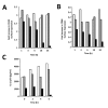
Red blood cell components did not affect maturation of dendritic cells. (A, B) To assess the percentage of phagocytosis, uninfected red blood cells (RBC), P. yoelii-infected RBC (iRBC) at the schizont stage and RBC treated with ionomycin (A) or different stages of P. yoelii-infected RBC (B) were labeled with the fluorescent dye CellTrace™ Far Red DDAO-SE, and incubated for 4 h with DC, before analysis by flow cytometry. The grey curve represents control DC that were not incubated with RBC. (C) CD86 expression analyzed by flow cytometry of DC incubated for 24 h with RBC, iRBC, Ionomycin-treated RBC (iono-RBC) and mouse hemoglobin (Hb). (D) CD86 expression analyzed by flow cytometry of DC incubated for 24 h with RBC, iRBC, Ionomycin-treated RBC (iono-RBC) and mouse hemoglobin (Hb), followed by another 18 h incubation with LPS (black bars) or PBS (white bars). Data correspond to the average of triplicate samples ± standard deviation and figures are representative of one out of three independent experiments.
Figure 7
Figure 7
Inhibition of LPS-induced maturation by P. yoelii-infected red blood cells (iRBC) is established between 6 and 18 h. To investigate the kinetics of the iRBC-induced inhibition of DC maturation, DC were pre-incubated for different times (1 to 24 h) with no RBC (white bars), uninfected RBC at 30:1 (grey bars) or iRBC at 30:1 (black bars), and then pulsed with LPS for 18 h. Cells were then analyzed by flow cytometry for expression of CD40 (A) and CD86 (B). To characterize the kinetics of the iRBC-induced inhibition of cytokine production, DC were pre-incubated for different times (0 to 24 h) with uninfected RBC at 30:1 (white bars) or iRBC at 10:1 (grey bars) or 30:1 (black bars), and then pulsed with LPS 1 μg/ml. Twenty-four hours after addition of LPS, supernatants of DC cultures were collected and IL-12p70 was quantified by ELISA. Data shows a representative experiment out of two independent experiments.
Figure 8
Figure 8
P. yoelii-infected red blood cells inhibit maturation of dendritic cells induced by stimulation of different Toll-like Receptors. To address the effect of P. yoelii-infected RBC (iRBC) on DC maturation induced by activation of different TLRs, DC were pre-incubated for 24 h with no RBC(white bars), uninfected RBC(gray bars) or iRBC(black bars) at both 10:1(solid bars) and 30:1(striped bars) ratios, and then pulsed with the following ligands of TLRs for an additional 18 hours: LPS 1 μg/ml, CpG-ODN 1 μg/ml, Zymosan 50 μg/ml, PolyI:C 25 μg/ml, flagellin 0.1 μg/ml and loxoribin 0.5 μM. DC were then analyzed by flow cytometry for expression of CD86. Data correspond to the average of triplicate samples ± standard deviation and figures are representative of one out of three independent experiments.

Similar articles

Cited by

References

    1. Good MF, Xu H, Wykes M, Engwerda CR. Development and regulation of cell-mediated immune responses to the blood stages of malaria: implications for vaccine research. Annu Rev Immunol. 2005;23:69–99. doi: 10.1146/annurev.immunol.23.021704.115638. - DOI - PubMed
    1. Guermonprez P, Valladeau J, Zitvogel L, Thery C, Amigorena S. Antigen presentation and T cell stimulation by dendritic cells. Annu Rev Immunol. 2002;20:621–667. doi: 10.1146/annurev.immunol.20.100301.064828. - DOI - PubMed
    1. Blander JM. Coupling Toll-like receptor signaling with phagocytosis: potentiation of antigen presentation. Trends Immunol. 2007;28:19–25. doi: 10.1016/j.it.2006.11.001. - DOI - PubMed
    1. Kool M, Petrilli V, De Smedt T, Rolaz A, Hammad H, van Nimwegen M, Bergen IM, Castillo R, Lambrecht BN, Tschopp J. Cutting edge: alum adjuvant stimulates inflammatory dendritic cells through activation of the NALP3 inflammasome. J Immunol. 2008;181:3755–3759. - PubMed
    1. Franchi L, Eigenbrod T, Munoz-Planillo R, Nunez G. The inflammasome: a caspase-1-activation platform that regulates immune responses and disease pathogenesis. Nat Immunol. 2009;10:241–247. doi: 10.1038/ni.1703. - DOI - PMC - PubMed

Publication types

MeSH terms