. 2010 Jun 15;401(2):217-27.
doi: 10.1016/j.ab.2010.02.030. Epub 2010 Mar 1.

Maximum yields of microsomal-type membranes from small amounts of plant material without requiring ultracentrifugation

Affiliations

Maximum yields of microsomal-type membranes from small amounts of plant material without requiring ultracentrifugation

Lindy Abas et al. Anal Biochem. .

Abstract

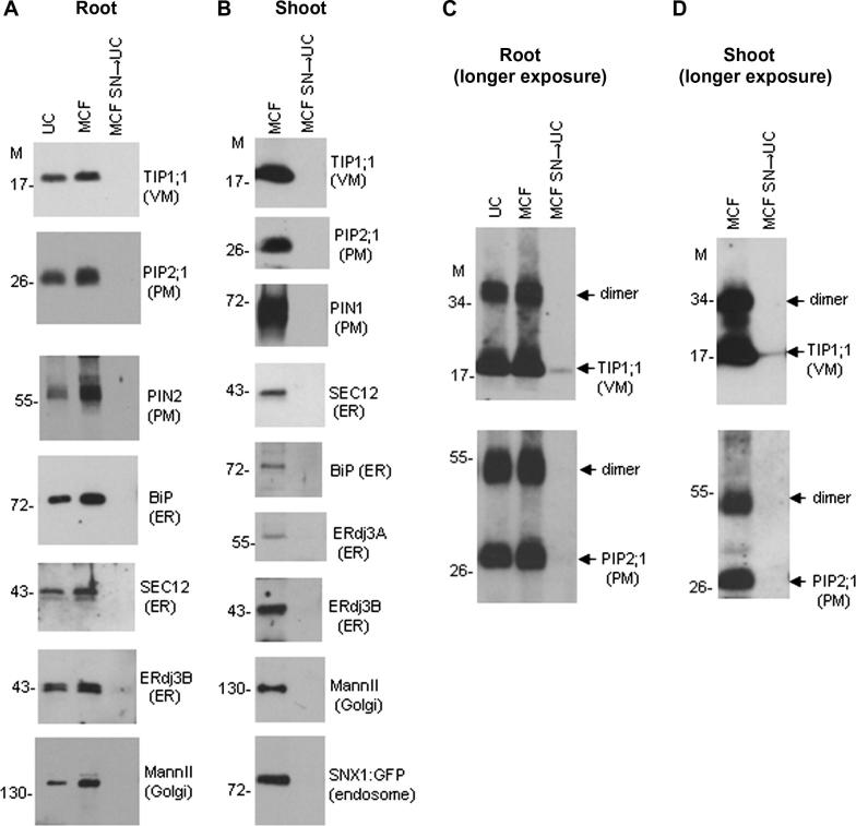
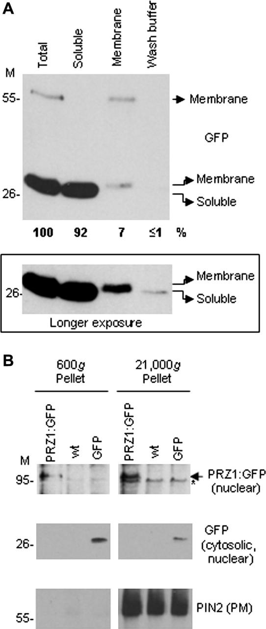
Isolation of a microsomal membrane fraction is a common procedure in studies involving membrane proteins. By conventional definition, microsomal membranes are collected by centrifugation of a postmitochondrial fraction at 100,000g in an ultracentrifuge, a method originally developed for large amounts of mammalian tissue. We present a method for isolating microsomal-type membranes from small amounts of Arabidopsis thaliana plant material that does not rely on ultracentrifugation but instead uses the lower relative centrifugal force (21,000g) of a microcentrifuge. We show that the 21,000g pellet is equivalent to that obtained at 100,000g and that it contains all of the membrane fractions expected in a conventional microsomal fraction. Our method incorporates specific manipulation of sample density throughout the procedure, with minimal preclearance, minimal volumes of extraction buffer, and minimal sedimentation pathlength. These features allow maximal membrane yields, enabling membrane isolation from limited amounts of material. We further demonstrate that conventional ultracentrifuge-based protocols give submaximal yields due to losses during early stages of the procedure; that is, extensive amounts of microsomal-type membranes can sediment prematurely during the typical preclearance steps. Our protocol avoids such losses, thereby ensuring maximal yield and a representative total membrane fraction. The principles of our method can be adapted for nonplant material.


Figures

Fig. 1
Fig. 1
Summary of the membrane isolation procedure: differences between a typical conventional UC protocol and our revised MCF protocol.
Fig. 2
Fig. 2
The MCF gives membrane yields equivalent to those of a UC. Shown is a Western blot analysis comparing the final membrane pellets prepared from identical material using either the MCF protocol or a UC-based protocol. To validate the efficiency of the MCF, the supernatant from the 21,000g spin was recentrifuged further at 100,000g so as to check for any remaining membranes (MCF SN → UC). Pellets were analyzed for the indicated marker proteins. Longer exposures for TIP1;1 and PIP2;1 were made to detect weak signals in the MCF SN → UC lanes (C and D). The observed SDS-stable dimers are expected for these proteins . All lanes are derived from 5-mg root (A and C) or 7-mg shoot (B and D). Molecular mass markers (M) are indicated (in kDa).
Fig. 3
Fig. 3
Conventional protocols give reduced yields of membrane proteins due to losses during preclearance. Shown is a Western blot analysis demonstrating the potential loss of desired membranes during typical conventional preclearance centrifugation steps. Crude homogenates were precleared either at 600g (minimal pre-clear) or further at 10,000g (A and B) or 2000g (C) to deplete mitochondria and/or chloroplasts (typical pre-clear), followed by final centrifugation at 100,000g or 21,000g. Pellets from preclearance (pre) and final centrifugation steps were analyzed for marker proteins. All lanes are derived from 5-mg root (A) or 7-mg shoot (B and C). Quantification (shown beneath the panels) is expressed as percentage of the total combined yield. Molecular mass markers (M, in kDa) and nonspecific bands (*) are indicated.
Fig. 4
Fig. 4
Minimal membrane loss during preclearance at 600g. Shown is a Western blot analysis demonstrating minimal loss of membranes by preclearing at 600g. Preclearance (600g) and final (21,000g) pellets from the MCF protocol were analyzed for the PM protein PIN2. All lanes are derived from 6-mg root. Molecular mass markers (M, in kDa) and nonspecific bands (*) are indicated.
Fig. 5
Fig. 5
A high-density EB reduces preclearance losses but must be diluted for complete sedimentation of membranes in the final centrifugation step. Shown is a Western blot analysis indicating that buffer density can be manipulated to control membrane sedimentation rates. (A) To compare the sedimentation of desired membranes during preclearance, root material was extracted using either a high-density EB (high, 25% sucrose) or a typical lower density EB (low, 8–10% sucrose). Homogenates were precleared at 10,000g (10 min), and these pellets analyzed for marker proteins. (B) To demonstrate that dilution is necessary for the final centrifugation step, shoot material was extracted with 25% sucrose EB and centrifuged undiluted at 21,000g (2 h) to collect membranes (initial pellet). The supernatant was then either diluted 2-fold (SN respun + dil) or left undiluted (SN respun undil) and recentrifuged at 21,000g (1 h). Pellets were analyzed for marker proteins. Quantification is expressed as percentage of low (A) or initial pellet (B). All lanes are derived from 5-mg root (A) or 7-mg shoot (B). Molecular mass markers (M) are indicated (in kDa).
Fig. 6
Fig. 6
Preservation of ER integrity and retention of peripheral membrane proteins in the membrane fraction. Shown is a Western blot analysis of membrane and soluble fractions following membrane isolation using the MCF protocol. Membranes were prepared from wild-type (A) or SNX1:GFP transgenic (B) seedlings. The 21,000g pellet (membrane) and supernatant (soluble) were analyzed for the indicated proteins. All lanes are derived from 2-mg seedlings. Quantification is expressed as percentage of the combined signals. Molecular mass markers (M) are indicated (in kDa).
Fig. 7
Fig. 7
A distinct form of membrane-associated GFP may be nuclear. Shown is a Western blot analysis of various fractions following membrane isolation. (A) Membranes were prepared from seedlings expressing free GFP. The total homogenate (total), 21,000g supernatant (soluble), 21,000g pellet (membrane), and wash (wash buffer) of the 21,000g pellet were analyzed for GFP. A longer exposure was made to detect the weak signal in the wash buffer (lower panel). Quantification is expressed as percentage of total. (B) Membranes were prepared from roots of wild-type (wt) seedlings or transgenic seedlings expressing GFP or PRZ1:GFP. The 600g and 21,000g pellets were analyzed for GFP or PIN2. All lanes are derived from 2-mg seedlings (A) or 6-mg root (B). Molecular mass markers (M, in kDa) and nonspecific bands (*) are indicated.

References

    1. Reid E., Williamson R. Centrifugation. Methods Enzymol. 1974;31:713–733. - PubMed
    1. Graham J. Isolation of subcellular organelles and membranes. In: Rickwood D., editor. Centrifugation: A Practical Approach. second ed. IRL Press; Oxford, UK: 1984. pp. 161–182.
    1. Ozols J. Preparation of membrane fractions. Methods Enzymol. 1990;182:225–235. - PubMed
    1. Robinson D.G., Hinz G., Oberbeck K. Isolation of endo- and plasma membranes. In: Harris N., Oparka K.J., editors. Plant Cell Biology: A Practical Approach. Oxford University Press; Oxford, UK: 1994. pp. 245–272.
    1. Dey P.M., Brownleader M.D., Harborne J.B. The plant, the cell, and its molecular components. In: Dey P.M., Harborne J.B., editors. Plant Biochemistry. Academic Press; San Diego: 1997. pp. 1–47.

Publication types

MeSH terms

LinkOut - more resources