Review
. 2010 Jul;49(7):1215-28.
doi: 10.1093/rheumatology/keq031. Epub 2010 Mar 1.

Transmembrane TNF-alpha: structure, function and interaction with anti-TNF agents

Affiliations
Review

Transmembrane TNF-alpha: structure, function and interaction with anti-TNF agents

Takahiko Horiuchi et al. Rheumatology (Oxford). 2010 Jul.

Abstract

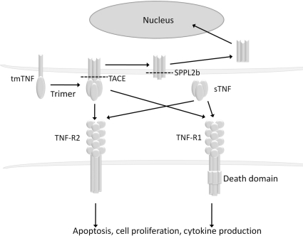
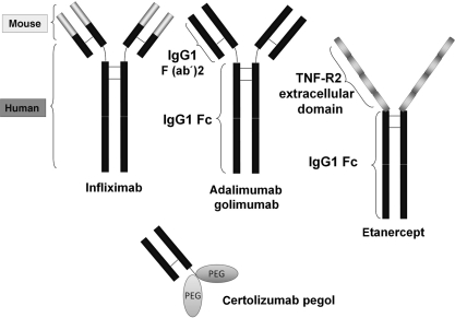
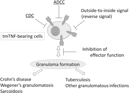
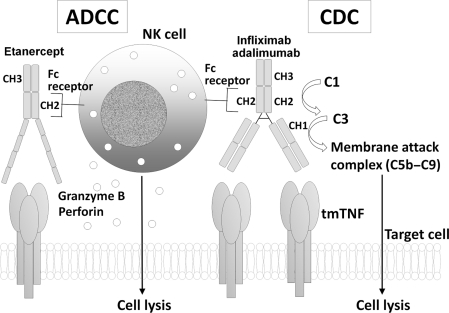
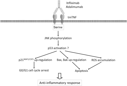
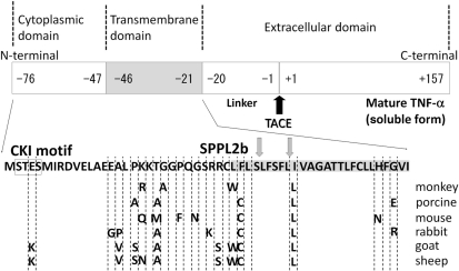
Transmembrane TNF-alpha, a precursor of the soluble form of TNF-alpha, is expressed on activated macrophages and lymphocytes as well as other cell types. After processing by TNF-alpha-converting enzyme (TACE), the soluble form of TNF-alpha is cleaved from transmembrane TNF-alpha and mediates its biological activities through binding to Types 1 and 2 TNF receptors (TNF-R1 and -R2) of remote tissues. Accumulating evidence suggests that not only soluble TNF-alpha, but also transmembrane TNF-alpha is involved in the inflammatory response. Transmembrane TNF-alpha acts as a bipolar molecule that transmits signals both as a ligand and as a receptor in a cell-to-cell contact fashion. Transmembrane TNF-alpha on TNF-alpha-producing cells binds to TNF-R1 and -R2, and transmits signals to the target cells as a ligand, whereas transmembrane TNF-alpha also acts as a receptor that transmits outside-to-inside (reverse) signals back to the cells after binding to its native receptors. Anti-TNF agents infliximab, adalimumab and etanercept bind to and neutralize soluble TNF-alpha, but exert different effects on transmembrane TNF-alpha-expressing cells (TNF-alpha-producing cells). In the clinical settings, these three anti-TNF agents are equally effective for RA, but etanercept is not effective for granulomatous diseases. Moreover, infliximab induces granulomatous infections more frequently than etanercept. Considering the important role of transmembrane TNF-alpha in granulomatous inflammation, reviewing the biology of transmembrane TNF-alpha and its interaction with anti-TNF agents will contribute to understanding the bases of differential clinical efficacy of these promising treatment modalities.


Figures

Fig. 1
Fig. 1
Biology of transmembrane TNF-α and soluble TNF-α. Transmembrane TNF-α is a precursor form of soluble TNF-α that is expressed on TNF-α-producing cells as a homotrimer. After processing by TACE, soluble TNF-α is generated and binds to TNF-R1 or -R2. Transmembrane TNF-α also binds to TNF-R1 and -R2. Upon binding to TNF receptors, both transmembrane and soluble TNF-α mediate pleiotropic effects (apoptosis, cell proliferation and cytokine production). The remaining transmembrane TNF-α after cleavage with TACE is further processed by SPPL2b and the intracellular domain is translocated into the nucleus and is supposed to mediate cytokine production. tmTNF: transmembrane TNF-α; sTNF: soluble TNF-α.
Fig. 2
Fig. 2
Structures of anti-TNF agents. Infliximab is a mouse–human chimeric monoclonal anti-TNF antibody of IgG1 isotype. Adalimumab and golimumab are fully human IgG1 monoclonal anti-TNF antibodies. Etanercept is a fusion protein of the extracellular domain of TNF-R2 and the Fc region of IgG1. Certolizumab pegol is a PEGylated Fab′ fragment of humanized monoclonal anti-TNF antibody.
Fig. 3
Fig. 3
Inhibition of TNF-α-bearing cells by anti-TNF agents. Transmembrane TNF-α plays an important role in granuloma formation, which is essential for the development of granulomatous diseases such as Crohn’s disease, and the host defence against tuberculosis. There are at least four distinct mechanisms for the inhibition of TNF-α-bearing cells by anti-TNF agents: (i) inhibition of transmembrane TNF-α-mediated effector function, (ii) destruction of TNF-α-bearing cells by CDC, (iii) destruction of TNF-α-bearing cells by ADCC and (iv) destruction of TNF-α-bearing cells by outside-to-inside signal (reverse signal).
Fig. 4
Fig. 4
CDC and ADCC by anti-TNF agents. Infliximab, adalimumab and etanercept commonly possess the Fc portion of IgG1, whose CH2 domain activates complement C1. Activation of C1 leads to complement C3 activation and subsequent formation of a membrane attack complex (C5b–C9) and lysis of the target cells. However, etanercept does not carry the CH1 domain of IgG1 which is important for the activation of C3. Infliximab, adalimumab and etanercept carry CH2 and CH3 domains of the Fc domain of IgG1 that mediate the binding to Fc receptors, which culminates in granzyme B and perforin release from NK cells and lysis of the target cells.
Fig. 5
Fig. 5
Outside-to-inside signal by adalimumab and infliximab. This is a novel mechanism for the inhibition of transmembrane TNF-α-bearing cells by anti-TNF antibodies. In the absence of NK cells or complement, adalimumab or infliximab induces G0/G1 cell cycle arrest and apoptosis, which inhibits TNF-α-producing cells and leads to an anti-inflammatory response. A number of molecules (p21WAF1/CIP1, Bax, Bak and ROS) were involved in these intracellular signalling events through the intracellular domain of transmembrane TNF-α. These signalling molecules are supposed to be associated with p53 activation. Three serine residues in the intracellular domain of transmembrane TNF-α are essential for the activities. Bak and Bax are proapoptotic multidomain molecules; tmTNF: transmembrane TNF-α.
Fig. 6
Fig. 6
Structure of transmembrane TNF-α. Transmembrane TNF-α is a type II polypeptide composed of a extracellular domain (177 amino acid residues), a transmembrane domain (26 amino acid residues, shaded) and an intracellular domain (30 amino acid residues). Mature TNF-α (soluble TNF-α) of 157 amino acid residues is cleaved from transmembrane TNF-α by TACE (black arrow). The remaining part is further cleaved by SPPL2b in the transmembrane domain (two grey arrows), and the intracellular domain is translocated into the nucleus to possibly modulate gene expression of the TNF-α-bearing cells. The intracellular domain contains CKI motif (boxed) and three serine residues. These serine residues are conserved among different species and are essential for the outside-to-inside signal transmitted by transmembrane TNF-α upon binding to anti-TNF antibody. Amino acid residues are shown in the one-letter code. The transmembrane domain of transmembrane TNF-α is shaded.

Similar articles

Cited by

References

    1. Feldmann M, Maini RN. Anti-TNF alpha therapy of rheumatoid arthritis: what have we learned? Annu Rev Immunol. 2001;19:163–96. - PubMed
    1. Bradley JR. TNF-mediated inflammatory disease. J Pathol. 2008;214:149–60. - PubMed
    1. Perez C, Albert I, DeFay K, Zachariades N, Gooding L, Kriegler M. A nonsecretable cell surface mutant of tumor necrosis factor (TNF) kills by cell-to-cell contact. Cell. 1990;63:251–8. - PubMed
    1. Georgopoulos S, Plows D, Kollias G. Transmembrane TNF is sufficient to induce localized tissue toxicity and chronic inflammatory arthritis in transgenic mice. J Inflamm. 1996;46:86–97. - PubMed
    1. Alexopoulou L, Pasparakis M, Kollias G. A murine transmembrane tumor necrosis factor (TNF) transgene induces arthritis by cooperative p55/p75 TNF receptor signaling. Eur J Immunol. 1997;27:2588–92. - PubMed

Publication types

MeSH terms