. 2010 Mar;92(3):599-608.
doi: 10.2106/JBJS.I.00425.

In vivo effects of single intra-articular injection of 0.5% bupivacaine on articular cartilage

Affiliations

In vivo effects of single intra-articular injection of 0.5% bupivacaine on articular cartilage

Constance R Chu et al. J Bone Joint Surg Am. 2010 Mar.

Abstract

Background: Single intra-articular injections of local anesthetics are commonly used clinically. Recent in vitro studies have demonstrated chondrotoxic effects of local anesthetics, with the greatest emphasis on bupivacaine toxicity. This in vivo study was conducted to determine whether a single intra-articular injection of 0.5% bupivacaine results in chondrocyte morbidity and rapid chondrolysis.

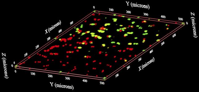
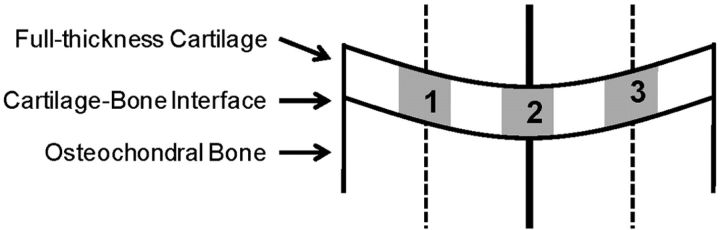
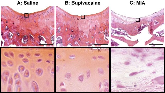
Methods: Forty-eight Sprague-Dawley rats received a 100-microL injection of sterile 0.9% saline solution (negative control) into one stifle joint and 100 microL of either preservative-free 0.5% bupivacaine (experimental group) or 0.6 mg/mL monoiodoacetate (positive control) into the contralateral joint. The rats were killed at one week, four weeks, twelve weeks, or six months. Live and dead cells were quantified with use of three-dimensional confocal reconstructions of fluorescent-stained tissues at standardized locations on the distal part of the femur. Histological findings were graded with use of a modified Mankin score, and cell density was quantified with use of custom image-analysis software.

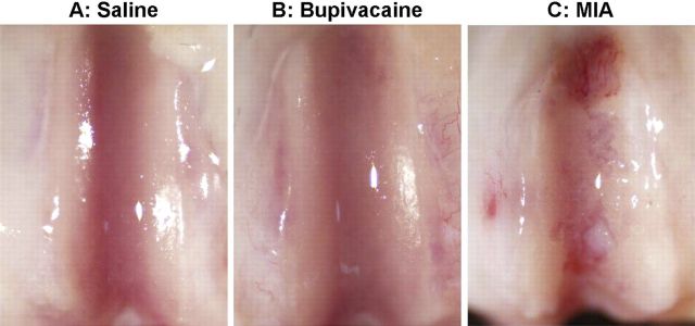
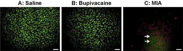
Results: In the specimens injected with bupivacaine, the chondral surfaces remained intact as seen with gross and histological examination. No differences in superficial chondrocyte viability or modified Mankin scores were observed between the saline-solution and bupivacaine groups at any location or time point (p > 0.05). Quantitative histological analysis of the bupivacaine-treated knees at six months revealed an up to 50% reduction in chondrocyte density compared with that of the saline-solution-treated knees (p < or = 0.01). Monoiodoacetate injection resulted in death of up to 87% of the superficial chondrocyte cells at one week and chondrolysis at six months. Despite severe histological abnormalities by four weeks after monoiodoacetate injection, cartilage injury was not evident on gross inspection until six months.

Conclusions: This in vivo study showing reduced chondrocyte density without cartilage tissue loss six months after a single intra-articular injection of 0.5% bupivacaine suggests bupivacaine toxicity. The effects of bupivacaine were milder than those of an injection of 0.6% monoiodoacetate, which resulted in chondrolysis over the same time period.


Figures

Fig. 1
Fig. 1
Quantification of viable chondrocytes with use of confocal microscopy. Following fluorescent staining with propidium iodide (red, indicating dead cells) and CMFDA (green, indicating live cells), a serial z-axis image stack 720 × 540 μm was acquired from the articular surface of the center of the medial and lateral femoral condyles and the center of the trochlear groove to a depth of 25 μm. Image stacks were then reconstructed in three-dimensional space. The percent cell viability was counted in each three-dimensional image.
Fig. 2
Fig. 2
Computerized quantification of histological sections. Following segmentation of the articular cartilage with use of customized histomorphometric software, the center of the trochlea, the medial femoral condyle, or the lateral femoral condyle was identified on each section. A line was drawn at the center, perpendicular to the cartilage surface, dividing the image into two halves (solid line). Each half section was again bisected, and a second line was drawn (dashed lines). The software was then used to generate three full-thickness cartilage regions of interest, centered on each line, of approximately equal size (∼300 μm wide). The mean cell density was then calculated within each region of interest, and the densities were averaged for each histological section.
Fig. 3
Fig. 3
Gross assessment of chondral surfaces. The articular surfaces were smooth and glistening, with no visible surface irregularities, six months after a single intra-articular injection of normal saline solution (A) or 0.5% bupivacaine (B). In contrast, a single injection of 0.6% monoiodoacetate (MIA) (C) resulted in visible cartilage erosions.
Fig. 4
Fig. 4
Surface chondrocyte viability. No significant loss of surface chondrocyte viability was seen one week after a single intra-articular injection of normal saline solution (A) or 0.5% bupivacaine (B) (p > 0.05 for the comparison between the two groups). In contrast, a single injection of 0.6% monoiodoacetate (MIA) (C) resulted in up to 87% cell death in the femoral condyles and trochlear groove (p < 0.05 compared with the saline-solution group). Viability was assessed with red/green staining and examination with three-dimensional confocal microscopy (living cells stained green [CMFDA] and dead cells stained red [propidium iodide]). The arrows indicate empty lacunae, seen only in the monoiodoacetate group with confocal microscopy. Scale bar = 60 μm.
Fig. 5
Fig. 5
Histological appearance and surface chondrocyte viability following monoiodoacetate injection. No difference in the total modified Mankin scores was observed, at any of the study time points, between specimens given a single intra-articular injection of normal saline solution and those given a single intra-articular injection of 0.5% bupivacaine. Injection of 0.6% monoiodoacetate resulted in higher total modified Mankin scores, indicative of increasing degeneration, by four weeks (p < 0.01 compared with those in the saline-solution group), with the highest scores obtained at six months (p < 0.001 compared with those in the saline-solution group). Comparison of the results of the viability imaging and histological assessment of the monoiodoacetate-injected specimens demonstrated consistent and complementary information. A: One week after monoiodoacetate injection, viability assessment showed substantial chondrocyte necrosis despite a normal histological appearance. B: At four weeks, both viability imaging and histological examination showed acellular tissue with empty lacunae and chondrocyte drop-out from superficial to deep zones. C: At twelve weeks, partial cellular repopulation was observed, especially in the superficial zone, with both viability imaging and histological analysis. D: At six months, scattered live and dead cells were seen with viability imaging. Histological examination revealed full-thickness cartilage loss and fibrosis. Scale bars on fluorescent stereomicroscopy images = 40 μm. Scale bars on histological images = 100 μm.
Fig. 6
Fig. 6
Chondrocyte density. Quantitative image analysis of histological specimens showed that chondrocyte density in specimens injected with a single intra-articular injection of 0.5% bupivacaine, while similar to that in the saline-solution group at one week after injection, was 50% less than that in the saline-solution group at six months after injection (p = 0.017). Histological images show the density of chondrocytes in the saline-solution-injected specimen (A) to be higher than that in the bupivacaine-injected specimen (B) at six months. Six months after injection with 0.6% monoiodoacetate (MIA) (C), replacement with fibrous repair tissue can be seen. Scale bar = 250 μm.

References

    1. Chu CR Izzo NJ Coyle CH Papas NE Logar A. The in vitro effects of bupivacaine on articular chondrocytes. J Bone Joint Surg Br. 2008;90:814-20. - PMC - PubMed
    1. Chu CR Izzo NJ Papas NE Fu FH. In vitro exposure to 0.5% bupivacaine is cytotoxic to bovine articular chondrocytes. Arthroscopy. 2006;22:693-9. - PubMed
    1. Dragoo JL Korotkova T Kanwar R Wood B. The effect of local anesthetics administered via pain pump on chondrocyte viability. Am J Sports Med. 2008;36:1484-8. - PubMed
    1. Gomoll AH Kang RW Williams JM Bach BR Cole BJ. Chondrolysis after continuous intra-articular bupivacaine infusion: an experimental model investigating chondrotoxicity in the rabbit shoulder. Arthroscopy. 2006;22:813-9. - PubMed
    1. Hansen BP Beck CL Beck EP Townsley RW. Postarthroscopic glenohumeral chondrolysis. Am J Sports Med. 2007;35:1628-34. - PubMed