. 2010 Mar;92(3):675-85.
doi: 10.2106/JBJS.H.01672.

Multipotential differentiation of human anulus fibrosus cells: an in vitro study

Affiliations

Multipotential differentiation of human anulus fibrosus cells: an in vitro study

Gang Feng et al. J Bone Joint Surg Am. 2010 Mar.

Abstract

Background: The existence of fibrocartilage, bone-like tissues, nerves, and blood vessels in the anulus fibrosus during intervertebral disc degeneration has been well documented. Migration of differentiated cells from outside the intervertebral disc has been hypothesized as a possible mechanism for the formation of these tissues. We hypothesized that the normal anulus fibrosus tissue contains multipotent progenitor cells, which are able to differentiate into cartilage and/or fibrocartilage cells, osteoblasts, neurons, and blood vessel cells.

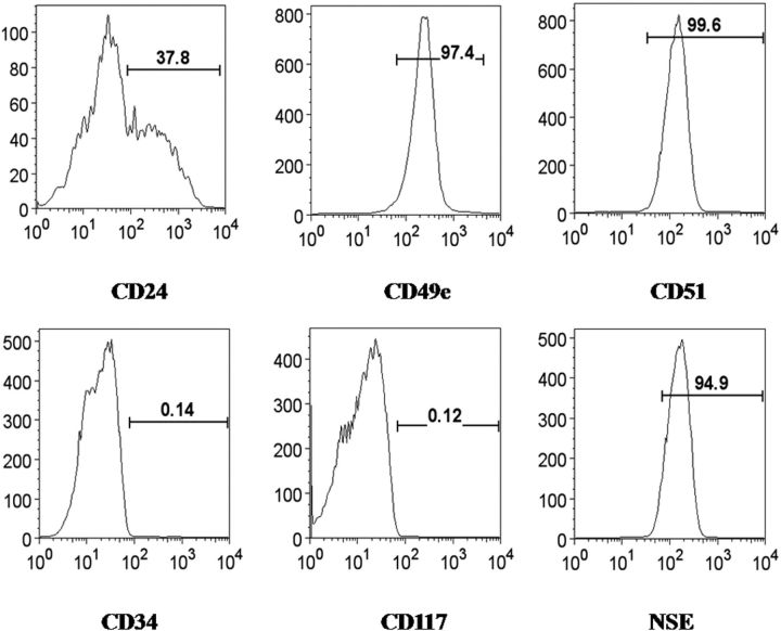
Methods: We isolated anulus fibrosus cells from the nondegenerative intervertebral discs of adolescent (thirteen to sixteen-year-old) patients with idiopathic scoliosis and cultured the cells in vitro in induction media containing different stimuli. Immunophenotypic analysis of cell surface markers was performed by flow cytometry. Expression of markers of adipogenesis, osteogenesis, chondrogenesis, neurogenesis, and differentiation into endothelial lineages was determined with use of immunostaining, cytohistological staining, and reverse transcription-polymerase chain reaction.

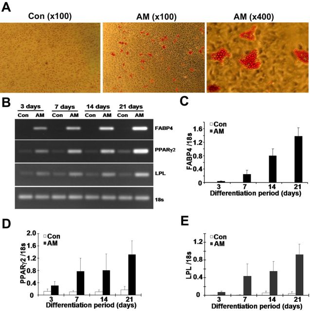
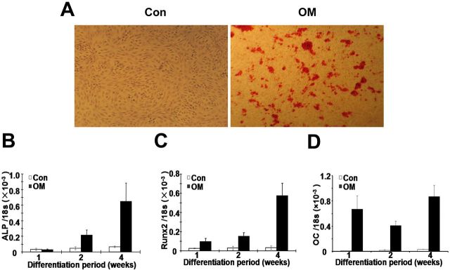
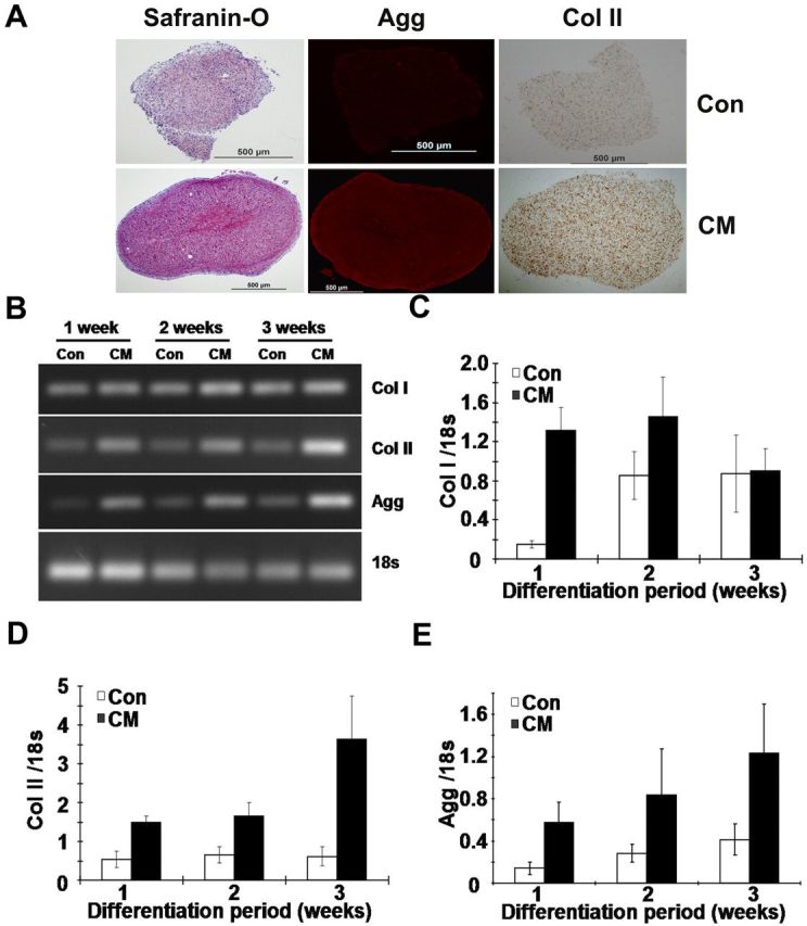
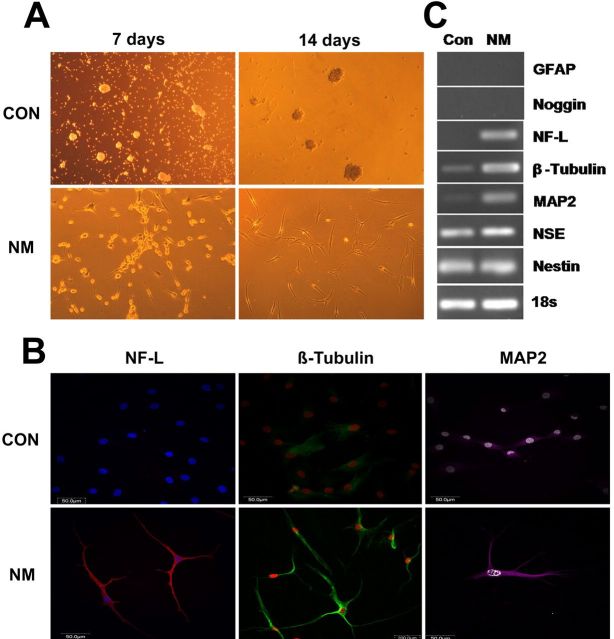
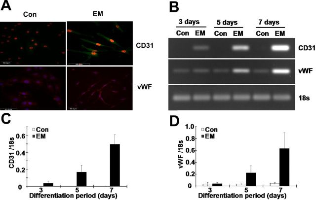
Results: Anulus fibrosus cells expressed several of the cell surface antigens that are sometimes associated with mesenchymal stem cells, including CD29, CD49e, CD51, CD73, CD90, CD105, CD166, CD184, and Stro-1, and two neuronal stem cell markers, nestin and neuron-specific enolase. Furthermore, varying the stimulants added to the induction media determined whether anulus fibrosus cells differentiated into adipocytes, osteoblasts, chondrocytes, neurons, or endothelial cells.

Conclusions: Anulus fibrosus cells isolated from nondegenerative intervertebral discs can differentiate into adipocytes, osteoblasts, chondrocytes, neurons, and endothelial cells in vitro.


Figures

Fig. 1
Fig. 1
Representative flow cytometry analysis of the expression of cell markers in cultured anulus fibrosus cells isolated from a human intervertebral disc. The cell markers CD29, CD49e, CD51, CD73, CD90, CD105, CD166, and CD184, as well as the neuronal markers nestin and neuron-specific enolase (NSE) were all expressed, whereas other markers were negative or partially positive. The horizontal axis represents the parameter's signal value in channel numbers, and the vertical axis represents the number of events per channel number. The number in each panel shows the percentage of cells expressing the specific cell marker.
Fig. 2
Fig. 2
Adipogenic differentiation of cultured human anulus fibrosus cells was induced with use of the commercial adipogenic supplements in five samples. (A) Oil-red-O staining showing formation of lipid droplets within induced cells. Reverse transcription-polymerase chain reaction (B) and real-time reverse transcription-polymerase chain reaction (C, D, and E) analysis of cellular mRNA levels of several adipogenesis-specific proteins, with 18S rRNA as an internal control. The gene expression levels of fatty acid binding protein 4 (FABP4) (C), peroxisome proliferator-activated receptor-gamma 2 (PPARγ2) (D), and lipoprotein lipase (LPL) (E) are increased in anulus fibrosus cells treated with adipogenic media (AM) compared with control cells (Con) cultured in basal media. Error bars represent the standard deviation.
Fig. 3
Fig. 3
Osteogenic differentiation of cultured human anulus fibrosus cells was induced with use of the osteogenic supplements. Once cells had reached 100% confluence, they were induced for up to four weeks and then were stained with alizarin red S or assayed for gene expression. Mineralization in induced cells is revealed by alizarin red S (×40) (A). Real-time reverse transcription-polymerase chain reaction analysis of osteogenesis-specific genes alkaline phosphatase (ALP) (B), runt-related transcription factor 2 (Runx2) (C), and osteocalcin (OC) (D) was performed, with 18S rRNA as an internal control (Con). The expression of all three genes was upregulated following the induction protocol in five samples. Error bars represent the standard deviation. OM = osteogenic media.
Fig. 4
Fig. 4
Chondrogenic differentiation of human anulus fibrosus cells treated with the chondrogenic supplements. Cells were induced for three weeks in a pellet cell culture system, and then the expression of several extracellular matrix proteins was assayed by cytochemical and reverse transcription-polymerase chain reaction analysis. Safranin-O staining of proteoglycan and immunocytochemical staining of aggrecan (Agg) and type-II collagen (Col II) (A). Reverse transcription-polymerase chain reaction (B) and real-time reverse transcription-polymerase chain reaction analysis of type-I (Col I) (C) and type-II collagen (D) and aggrecan (E) mRNA levels, with 18S rRNA as an internal control (Con). Expression of type-II collagen and aggrecan was substantially enhanced after chondrogenic induction in five samples. Error bars represent the standard deviation. CM = chondrogenic media.
Fig. 5
Fig. 5
Neurogenic differentiation of human anulus fibrosus cells by a sphere culture method with use of the NeuroCult NS-A Proliferation Kit and NeuroCult NS-A Differentiation Kit. After induction, several neuronal markers were examined by immunofluorescent staining and reverse transcription-polymerase chain reaction analysis. A: Cell morphology under a light microscope (×100). Cells displayed a neuron-like appearance after they were induced for fourteen days. B: Immunofluorescent staining of neurofilament light chain (NF-L), β-tubulin, and microtubule-associated protein 2 (MAP2). C: Reverse transcription-polymerase chain reaction analysis of glial fibrillary acidic protein (GFAP), noggin, neurofilament light chain, β-tubulin, microtubule-associated protein 2, neuron-specific enolase (NSE), and nestin mRNA levels, with 18S rRNA as an internal control (Con). Expression of β-tubulin, microtubule-associated protein 2, and neurofilament light chain was promoted after induction. NM = neurogenic media.
Fig. 6
Fig. 6
Endothelial lineage differentiation of cultured human anulus fibrosus cells. Cells were incubated for up to seven days in Endothelial Cell Growth Medium MV 2, and then the expression of endothelial-specific genes was examined by immunofluorescent staining and reverse transcription-polymerase chain reaction. Cell morphology and immunofluorescent staining of CD31 and von Willebrand factor (vWF) (A). Reverse transcription-polymerase chain reaction (B) and real-time reverse transcription-polymerase chain reaction analysis of the levels of the mRNA for CD31 (C) and vWF (D) mRNA, with 18S rRNA as an internal control (Con). The expression of both genes was upregulated following induction in six samples. EM = endothelial media.

References

    1. Diamond S Borenstein D. Chronic low back pain in a working-age adult. Best Pract Res Clin Rheumatol. 2006;20:707-20. - PubMed
    1. Luo X Pietrobon R Sun SX Liu GG Hey L. Estimates and patterns of direct health care expenditures among individuals with back pain in the United States. Spine (Phila Pa 1976). 2004;29:79-86. - PubMed
    1. Battié MC Videman T Levalahti E Gill K Kaprio J. Heritability of low back pain and the role of disc degeneration. Pain. 2007;131:272-80. - PubMed
    1. Andersson GB. Epidemiological features of chronic low-back pain. Lancet. 1999;354:581-5. - PubMed
    1. Walker MH Anderson DG. Molecular basis of intervertebral disc degeneration. Spine J. 2004;4:158S-66S. - PubMed