

doi: 10.1101/gad.1864410.
KDM7 is a dual demethylase for histone H3 Lys 9 and Lys 27 and functions in brain development
Affiliations
- PMID: 20194436
- PMCID: PMC2827838
- DOI: 10.1101/gad.1864410
Item in Clipboard
KDM7 is a dual demethylase for histone H3 Lys 9 and Lys 27 and functions in brain development
Genes Dev.
.
Abstract
Methylation of histone H3 Lys 9 and Lys 27 (H3K9 and H3K27) is associated with transcriptional silencing. Here we show that KDM7, a JmjC domain-containing protein, catalyzes demethylation of both mono- or dimethylated H3K9 and H3K27. Inhibition of KDM7 orthologs in zebrafish resulted in developmental brain defects. KDM7 interacts with the follistatin gene locus, and KDM7 depletion in mammalian neuronal cells suppressed follistatin gene transcription in association with increased levels of dimethylated H3K9 and H3K27. Our findings identify KDM7 as a dual demethylase for H3K9 and H3K27 that functions as an eraser of silencing marks on chromatin during brain development.
Figures
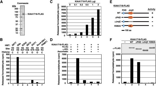
KIAA1718 is a histone demethylase that targets H3K9. (A, right lane) SDS–polyacrylamide gel electrophoresis with Coomassie blue staining of a C-terminally Flag-tagged recombinant KIAA1718 protein. Molecular size standards are shown in the left lane. (B) Histone demethylase activity of purified KIAA1718-Flag with various methylated histone substrates. The HMTs used to generate the various substrates and their sites of methylation are indicated. The methylated substrates were generated with the indicated forms of histone ([C] core histone octamer; [O] oligonucleosome) on the basis of the substrate preference of each HMT. The presented counts have been corrected for control counts. (C) Histone demethylase activity of the indicated amounts of KIAA1718-Flag. (D) Effects of removal of Fe(II), α-KG, or ascorbate from the reaction mixture on the histone demethylase activity of KIAA1718-Flag with G9a-methylated histone substrate. (E) Schematic representation of wild-type (WT) and mutant forms of KIAA1718 showing whether they are active (+) or inactive (−) as H3K9 demethylases. (F, bottom panel) Demethylase activity of purified wild-type or mutant forms of KIAA1718-Flag with G9a-methylated histone substrate. (Top panel) The similar amounts of KIAA1718-Flag proteins used in the demethylase assay are revealed by immunoblot analysis with antibodies to Flag (α-Flag).
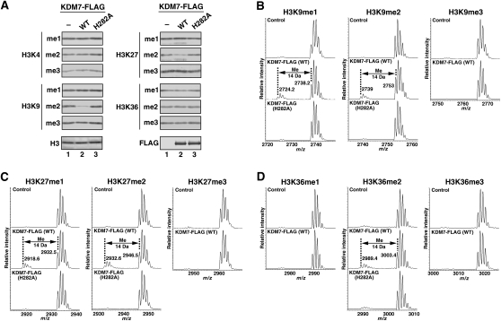
KDM7 is a histone demethylase specific for dimethylated or monomethylated H3K9 or H3K27. (A) Calf thymus core histones were incubated in the absence or presence of 5 μg of wild-type or H282A mutant forms of KDM7-Flag, after which histone demethylation was evaluated by immunoblot analysis with antibodies to specific modified histones, as indicated on the left. (B–D) Mass spectrometric analysis of the demethylase activity of 4 μg of wild-type or H282A mutant forms of KDM7-Flag with methylated H3K9, H3K27, or H3K36 peptide substrates. Numbers represent the masses of the peptide substrates and products.
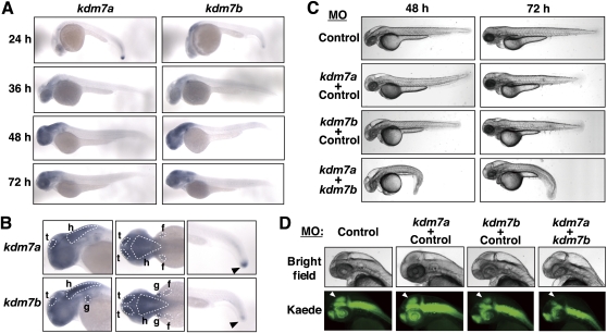
Zebrafish kdm7 genes are expressed predominantly in brain and are required for tectum development. (A) In situ hybridization of whole-mount zebrafish embryos at the indicated stages (hpf) with antisense kdm7a or kdm7b RNA probes. (B) In situ hybridization of whole-mount embryos at 48 hpf (left and middle panels) or 24 hpf (right panels) with antisense kdm7a or kdm7b RNA probes. Lowercase letters indicate the tectum (t), hindbrain (h), fin bud (f), and gill (g). Arrowheads indicate the tail bud. (C) The Tg(HuC:Kaede) embryos were injected at the one-cell stage with antisense MOs for kdm7a (5 ng) or kdm7b (5 ng) or with a control MO (5 or 10 ng for a total of 10 ng of MO) in the indicated combinations. The morphology of the embryos at the indicated times (hpf) was examined by bright-field microscopy. (D) Morphology of the head region of embryos at 48 hpf that were injected with MOs as in C. (Bottom panels) Neurons expressing Kaede under the control of the HuC gene promoter were visualized by fluorescence microscopy. Arrowheads indicate the tectum or presumptive tectal region. Bright-field images are shown in the top panels.
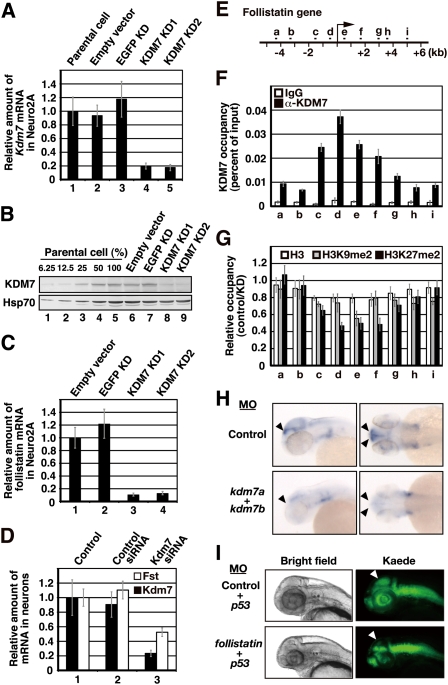
KDM7 directly regulates transcription and both H3K9me2 and H3K27me2 levels of the mouse follistatin gene. (A) Quantitative RT–PCR analysis of Kdm7 mRNA in Neuro2A cell lines stably transfected with vectors for one of two KDM7 shRNAs (KD1 or KD2), with a vector for a control (EGFP) shRNA, or with the empty vector. The amount of Kdm7 mRNA was normalized by that of Gapdh mRNA, and the normalized values are presented relative to that for the parental cells. Data are means ± SD. (B) Immunoblot analysis of KDM7 and Hsp70 (loading control) in the cell lines described in A. (C) Quantitative RT–PCR analysis of follistatin mRNA in the cell lines described in A. The amount of follistatin mRNA was normalized by that of Gapdh mRNA, and the normalized values are presented relative to that for the cells transfected with the empty vector. Data are means ± SD. (D) Quantitative RT–PCR analysis of Kdm7 and follistatin (Fst) mRNAs in primary cultured mouse neurons treated with control or Kdm7 siRNAs. The amounts of Kdm7 and follistatin mRNAs were normalized by that of Gapdh mRNA, and the normalized values are presented relative to that for control cells. Data are means ± SD. (E) Schematic representation of the mouse follistatin genomic locus. The region from a to i was analyzed by ChIP experiments. The transcription start site is indicated by the arrow. (F,G) ChIP analysis of the relative occupancy of the sites in the follistatin genomic region indicated in E with KMD7 (F), as well as with H3 (white bars), H3K9me2 (gray bars), and H3K27me2 (black bars) (G). The analysis was performed with cells stably transfected with the vector for EGFP shRNA (control) and with cells stably expressing the KD1 shRNA for KDM7 (KD), and the results are presented as the percent of input for control cells (F) or the control/KD ratio (G). All data are means ± SD. (H) In situ hybridization of whole-mount zebrafish embryos at 48 hpf with an antisense follistatin RNA probe. Arrowheads indicate regions expressing the follistatin gene in embryos injected at the one-cell stage with antisense MOs for kdm7a (5 ng) and kdm7b (5 ng) or with a control MO (10 ng). (I) The Tg(HuC:Kaede) embryos were injected at the one-cell stage with antisense MOs for follistatin (5 ng) or p53 (5 ng) genes or with a control MO (5 ng) in the indicated combinations. The morphology of the embryos at 48 hpf was examined by bright-field (left panels) or fluorescence (right panels) microscopy. Arrowheads indicate the tectum or presumptive tectal region.
References
-
- Allis CD, Berger SL, Cote J, Dent S, Jenuwien T, Kouzarides T, Pillus L, Reinberg D, Shi Y, Shiekhattar R, et al. New nomenclature for chromatin-modifying enzymes. Cell. 2007;131:633–636. - PubMed
-
- Bannister AJ, Schneider R, Kouzarides T. Histone methylation: Dynamic or static? Cell. 2002;109:801–806. - PubMed
-
- Hasenpusch-Theil K, Chadwick BP, Theil T, Heath SK, Wilkinson DG, Frischauf AM. PHF2, a novel PHD finger gene located on human chromosome 9q22. Mamm Genome. 1999;10:294–298. - PubMed
Publication types
MeSH terms
Substances
LinkOut - more resources
Full Text Sources
Molecular Biology Databases
Research Materials