. 2010 Mar 1;24(5):438-42.
doi: 10.1101/gad.1884910.

A degron created by SMN2 exon 7 skipping is a principal contributor to spinal muscular atrophy severity

Affiliations

A degron created by SMN2 exon 7 skipping is a principal contributor to spinal muscular atrophy severity

Sungchan Cho et al. Genes Dev. .

Abstract

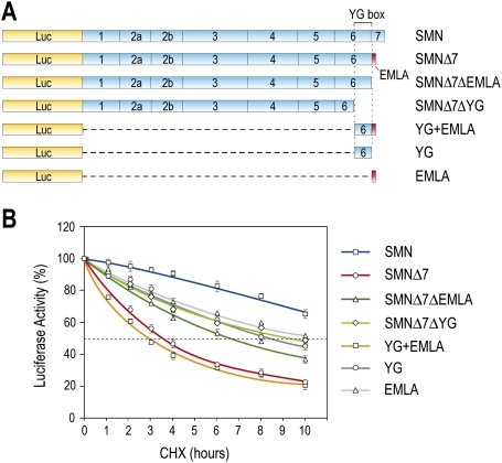
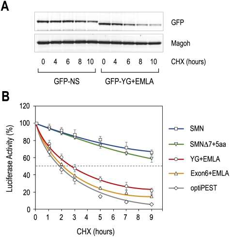
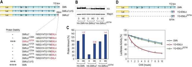
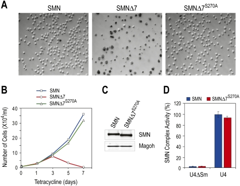
Spinal muscular atrophy (SMA) is caused by homozygous survival of motor neurons 1 (SMN1) gene deletions, leaving a duplicate gene, SMN2, as the sole source of SMN protein. However, most of the mRNA produced from SMN2 pre-mRNA is exon 7-skipped ( approximately 80%), resulting in a highly unstable and almost undetectable protein (SMNDelta7). We show that this splicing defect creates a potent degradation signal (degron; SMNDelta7-DEG) at SMNDelta7's C-terminal 15 amino acids. The S270A mutation inactivates SMNDelta7-DEG, generating a stable SMNDelta7 that rescues viability of SMN-deleted cells. These findings explain a key aspect of the SMA disease mechanism, and suggest new treatment approaches based on interference with SMNDelta7-DEG activity.


Figures

Figure 1.
Figure 1.
Delineation of YG + EMLA as a protein destabilization sequence in SMNΔ7. (A) Schematic diagram of Luc-fused SMN and a series of deletion constructs used for quantitative measurement of protein stability. Shown are SMN exon structures. YG box denotes the tyrosine/glycine (YG)-rich sequences in exon 6 of SMN. The EMLA sequence encoded by exon 8 is depicted by the red box at the C-terminal end of SMNΔ7. (B) 293T cells were transfected with plasmids expressing Luc-SMN, Luc-SMNΔ7, and the indicated deletion constructs. Forty-eight hours after transfection, the cells were treated with CHX (0.1 mg/mL) for various times as indicated, and then assayed for luciferase activity. Luc activity at each time point was calculated by comparison with those at time 0, which was set to 100%. Fifty percent activity is indicated by the gray dotted line. Error bars represent standard deviation (SD) from three independent experiments.
Figure 2.
Figure 2.
The C terminus of SMNΔ7, YG + EMLA, is a strong protein destabilizing signal (degron). (A) Plasmids expressing GFP-YG + EMLA or GFP-NS (nonspecific sequence) were transfected into 293T cells. Twenty-four hours after transfection, the cells were treated with CHX (0.1 mg/mL) for various times as indicated. GFP fusion proteins were detected by Western blot using anti-GFP antibody, and Magoh was used as a loading control. (B) Comparison of YG + EMLA and Exon6 + EMLA of SMNΔ7 with an optimized protein-destabilizing element (optiPEST). Shown also are Luc-SMN and Luc-SMNΔ7 containing an additional five amino acids at the C-terminal end (SMNΔ7 + 5aa). Luc activities were measured as in Figure 1. Error bars represent SDs from three independent experiments.
Figure 3.
Figure 3.
S270 is critical for the activity of the SMNΔ7-DEG through YG + EMLA. (A) Seven residues in the YG box were each mutated to alanine as indicated. All constructs had Luc fusions, and Luc activity was assayed as in Figure 1. (B) HA-tagged SMN, SMNΔ7, and SMNΔ7S270A were expressed in 293T cells for 24 h, and then cells were treated with DMSO (D) or 10 μM MG132 (MG) for 16 h. Fusion proteins were monitored by Western blot using an anti-HA tag antibody, and Magoh was used as a loading control. (C) HA-tagged proteins in B were quantified and compared with HA-SMN without MG132 treatment, which was set to 100%. The fold change of each fusion protein amount upon MG132 treatment is indicated in red above the column. (D) All constructs had Luc fusions, and Luc activity was assayed as in Figure 1. Error bars represent SDs from three independent experiments.
Figure 4.
Figure 4.
SMNΔ7S270A rescues SMN-deficient cells and is functional in snRNP assembly. (A) S5 cells were cultured in the presence of tetracycline (1 μg/mL) to deplete endogenous SMN, and were infected with retroviruses expressing SMN, SMNΔ7, or SMNΔ7S270A. One week after tetracycline addition, cells were stained with Trypan blue and visualized by DIC microscopy. (B) Cell growth as in A was measured by monitoring the number of live cells at the indicated time points following tetracycline addition. (C) Western blots of SMN protein in rescued cells (10 d after tetracycline addition). (D) Cytoplasmic extracts from rescued cells were assayed for snRNP assembly on U4 snRNA in vitro, using U4ΔSm RNA as a control.

Similar articles

Cited by

References

    1. Burnett BG, Crawford TO, Sumner CJ. Emerging treatment options for spinal muscular atrophy. Curr Treat Options Neurol. 2009a;11:90–101. - PubMed
    1. Burnett BG, Munoz E, Tandon A, Kwon DY, Sumner CJ, Fischbeck KH. Regulation of SMN protein stability. Mol Cell Biol. 2009b;29:1107–1115. - PMC - PubMed
    1. Chang HC, Hung WC, Chuang YJ, Jong YJ. Degradation of survival motor neuron (SMN) protein is mediated via the ubiquitin/proteasome pathway. Neurochem Int. 2004;45:1107–1112. - PubMed
    1. Cooper TA, Wan L, Dreyfuss G. RNA and disease. Cell. 2009;136:777–793. - PMC - PubMed
    1. Cusco I, Barcelo MJ, Rojas-Garcia R, Illa I, Gamez J, Cervera C, Pou A, Izquierdo G, Baiget M, Tizzano EF. SMN2 copy number predicts acute or chronic spinal muscular atrophy but does not account for intrafamilial variability in siblings. J Neurol. 2006;253:21–25. - PubMed

Publication types

Substances